File list
This special page shows all uploaded files.
| Date | Name | Thumbnail | Size | Description | Versions |
|---|---|---|---|---|---|
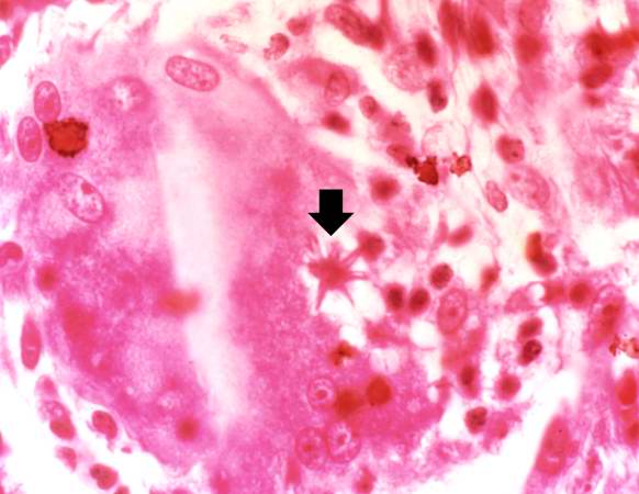
| 03:31, 19 August 2013 | IPLab3Sarcoidosis6.jpg (file) |  |
35 KB | This is a higher-power photomicrograph of an asteroid body (arrow) inside of a multinucleated giant cell. | 1 |
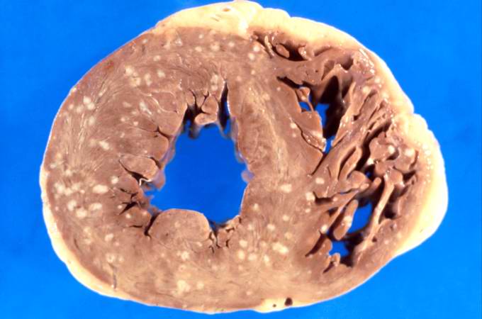
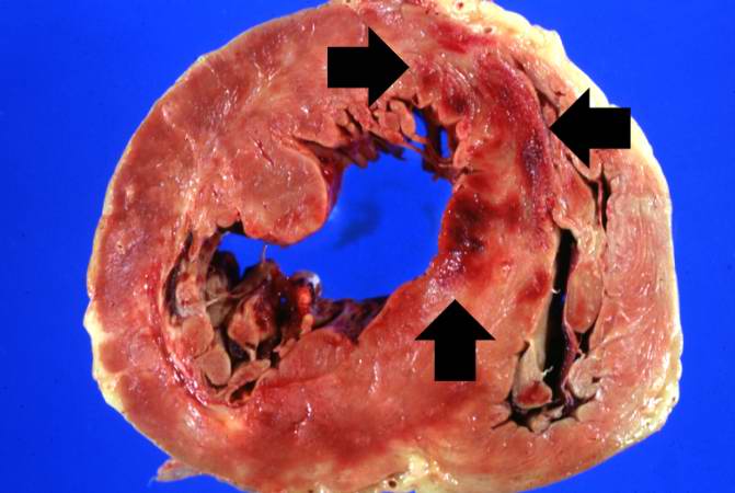
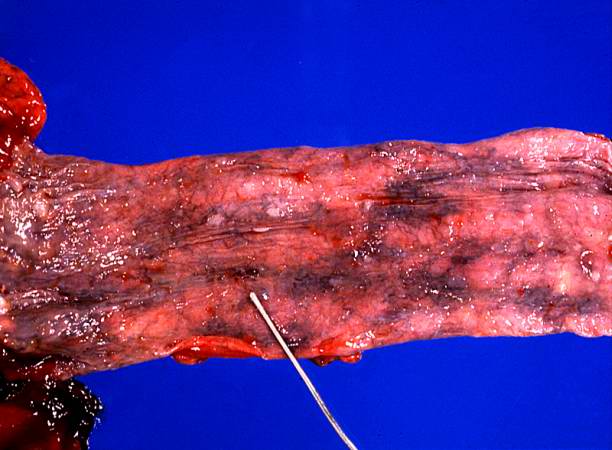
| 16:54, 19 August 2013 | IPLab4SepticEmboli2.jpg (file) |  |
35 KB | This is a gross photograph of myocardium with multiple embolic lesions scattered throughout the left and right ventricles. | 1 |
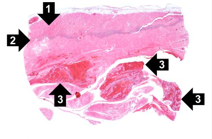
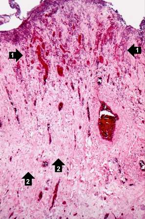
| 04:29, 19 August 2013 | IPLab3AcuteMyocardialInfarction1.jpg (file) |  |
36 KB | This is a low-power photomicrograph of infarcted heart. There is a layer of surviving myocardial tissue (1) along the epicardium and then a blue line (2) which represents the accumulation of inflammatory cells at the border of the infarct. There is thr... | 1 |
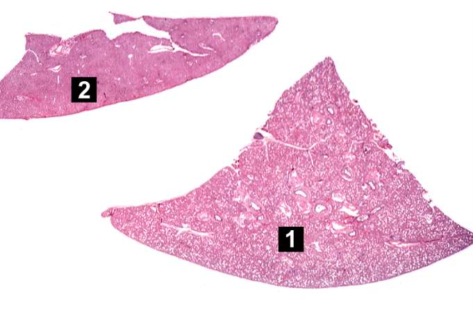
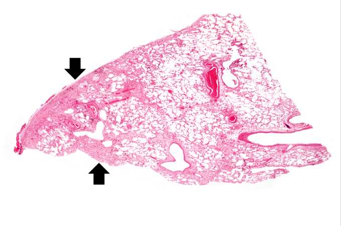
| 05:50, 21 August 2013 | IPLab13Hyaline2.jpg (file) |  |
36 KB | This is a low-power photomicrograph of a triangular-shaped section of lung (1) and an oblong section of liver (2). The lack of open air spaces in this neonatal lung indicates its immaturity. | 1 |
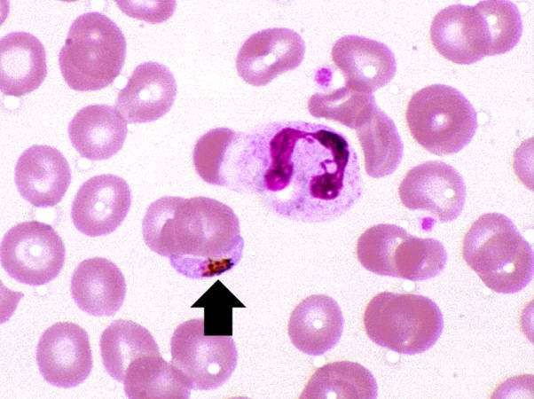
| 04:54, 21 August 2013 | IPLab11Malaria2.jpg (file) |  |
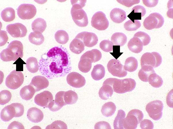
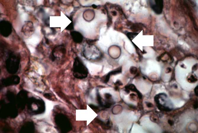
36 KB | This is another high power photomicrograph of a thin smear of blood from this patient. There is a single eosinophil in this smear along with several RBCs containing ring stage trophozoites (arrows). | 1 |
| 04:37, 19 August 2013 | IPLab3HealedMyocardialInfarction5.jpg (file) |  |
36 KB | This is a high-power photomicrograph of a different region of this healed MI. Note the chronic inflammatory reaction (arrows) in this region suggesting that there had been ischemic injury to this area within the last several weeks to months. | 1 |
| 02:37, 21 August 2013 | IPLab8HSVEncephalitis12.jpg (file) |  |
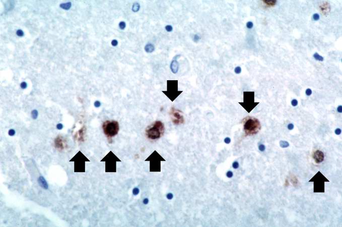
36 KB | This is a photomicrograph of a brain section stained with an antibody against herpes simplex. Even at this magnification, it is easy to pick out cells that are positive for the virus (arrows). | 1 |
| 03:56, 21 August 2013 | IPLab9Clostridium1.jpg (file) |  |
36 KB | This gross photograph of the lower extremity was taken at autopsy. Notice the swelling and the area of the primary infection (arrow). | 1 |
| 01:17, 16 August 2013 | IPLab1FatNecrosis1.jpg (file) |  |
36 KB | 1 | |
| 02:34, 21 August 2013 | IPLab8HSVEncephalitis2.jpg (file) |  |
36 KB | This is a closer view of the previous section of brain showing multiple small, punctate hemorrhages throughout the brain parenchyma (arrows). | 1 |
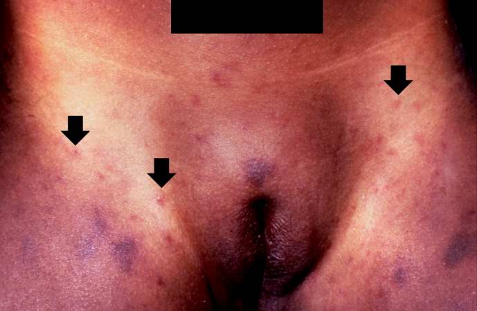
| 06:04, 21 August 2013 | IPLab13Meningococcemia2.jpg (file) |  |
36 KB | This is a closer view of the inguinal region taken at autopsy. The areas of hemorrhage include purpura and petechiae (arrows). | 1 |
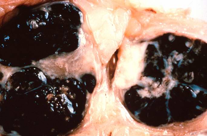
| 01:55, 21 August 2013 | IPLab7Melanoma2.jpg (file) |  |
36 KB | This is a gross photograph of lymph nodes almost entirely replaced by black pigment (melanin). | 1 |
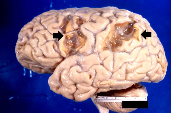
| 04:21, 19 August 2013 | IPLab3BrainInfarction1.jpg (file) |  |
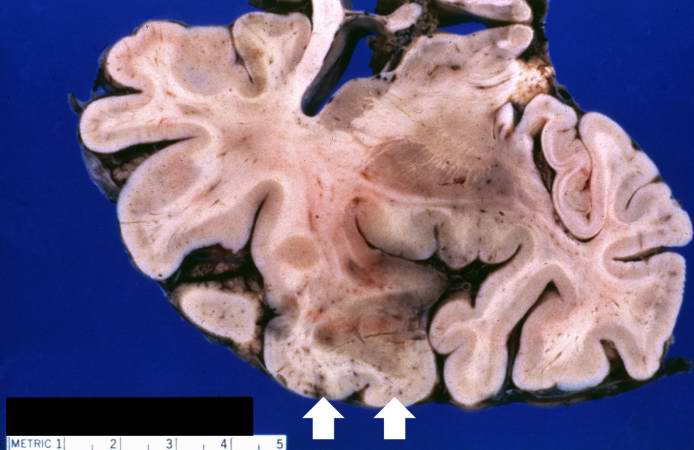
37 KB | This is a gross photograph of the brain which contains two areas of infarction (arrows). | 1 |
| 05:07, 21 August 2013 | IPLab11Chagas1.jpg (file) |  |
37 KB | This peripheral blood smear from the patient shows two trypomastigotes of Trypanosoma cruzi. | 1 |
| 04:55, 21 August 2013 | IPLab11Malaria5.jpg (file) |  |
37 KB | There is another example of a P. falciparum gametocyte (arrow) in this thin smear. There is a neutrophil in this field as well. | 1 |
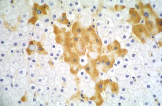
| 02:49, 21 August 2013 | IPLab8HBV8.jpg (file) |  |
37 KB | This higher-power photomicrograph of the previous section shows more clearly the HBsAg positive cells (arrows). Upon staining with H&E, these same cells exhibit a "ground glass" appearance, which is due to the accumulation of HBsAg in the hepatocyte cy... | 1 |
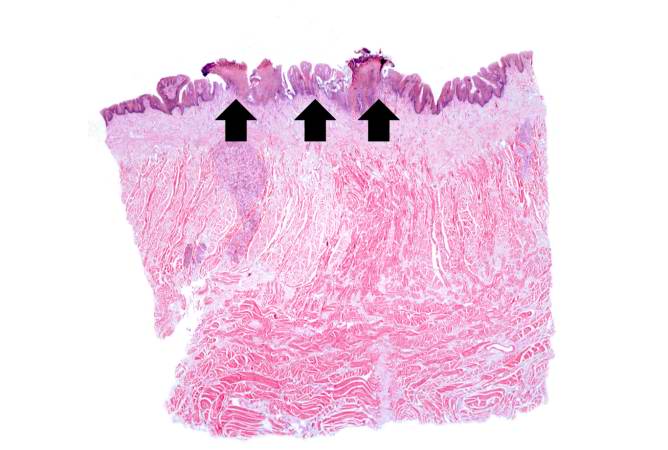
| 02:28, 21 August 2013 | IPLab8HSVGlossitis1.jpg (file) |  |
38 KB | This is a low-power photomicrograph showing a cross section of the tongue. There is an area along the surface of the tongue where the normal epithelium has been lost and there are areas of ulceration (arrows). | 1 |
| 05:30, 21 August 2013 | IPLab12RadiationChanges8.jpg (file) |  |
38 KB | This high-power photomicrograph of the wall of the ileum shows more examples of pleomorphic cells caused by radiation injury (arrows). | 1 |
| 04:58, 21 August 2013 | IPLab11Leishmaniasis5.jpg (file) |  |
38 KB | This is a high-power photomicrograph of an inflammatory cell containing cytoplasmic organisms (arrows). | 1 |
| 05:28, 21 August 2013 | IPLab12RadiationChanges3.jpg (file) |  |
38 KB | This is a higher-power photomicrograph showing the atrophied epithelium in the area of radiation injury. There are some epithelial cells deep within the mucosa (1). Note the dense fibrous connective tissue (2) within the wall of the ileum. | 1 |
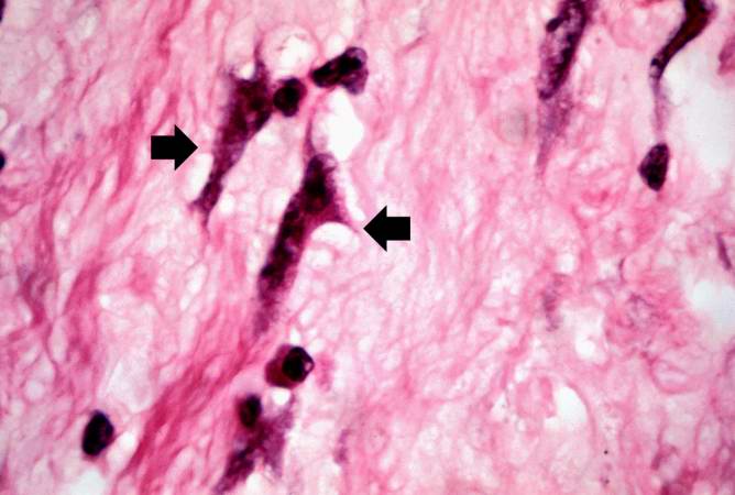
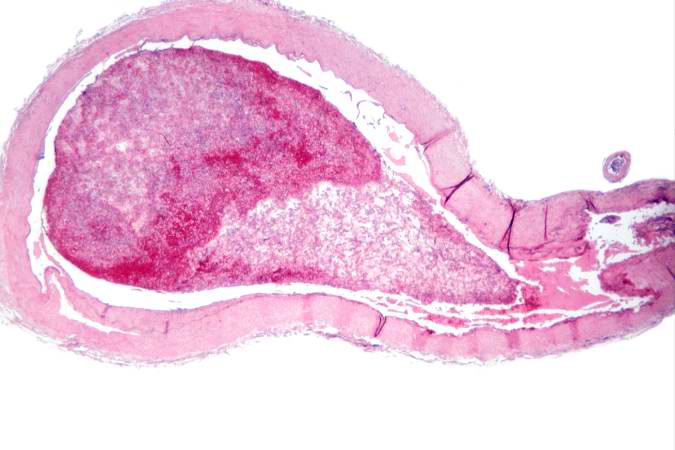
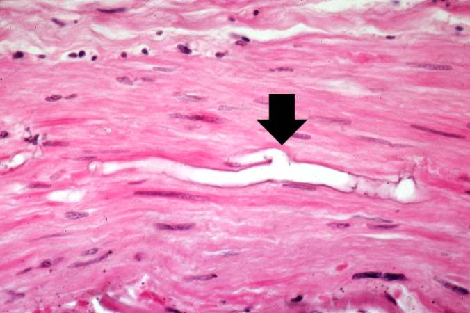
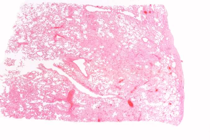
| 04:19, 21 August 2013 | IPLab10Mucor1.jpg (file) |  |
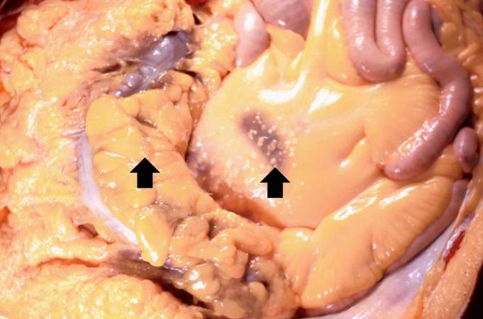
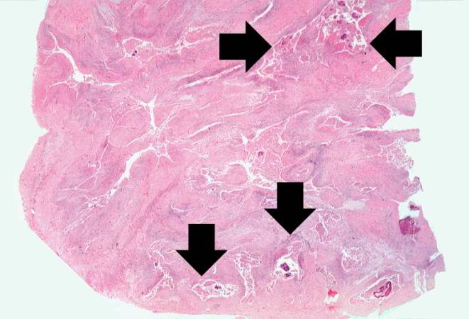
38 KB | This is a low-power photomicrograph of a section of carotid artery containing a mural thrombus. | 1 |
| 13:49, 15 August 2013 | IPLab1MyocardialInfarction1.jpg (file) |  |
38 KB | 1 | |
| 20:24, 20 August 2013 | IPLab6SenileAmyloidosis2.jpg (file) |  |
39 KB | This is a low power photomicrograph of the heart tissue from this case. At this magnification the structure looks relatively normal. | 1 |
| 04:12, 21 August 2013 | IPLab10Crypto9.jpg (file) |  |
39 KB | This higher-power photomicrograph of a cryptococcal organism shows more clearly the nucleus surrounded by the large extracellular capsule (arrows). | 1 |
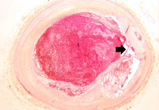
| 16:27, 19 August 2013 | IPLab4MuralThrombus2.jpg (file) |  |
39 KB | This is a low-power photomicrograph of the thrombus (1) attached to the myocardium (2). | 1 |
| 05:28, 21 August 2013 | IPLab12RadiationChanges4.jpg (file) |  |
39 KB | This is a high-power photomicrograph showing the atrophied epithelium in the area of radiation injury (1). Note the dense fibrous connective tissue within the wall of the ileum and the congested blood vessels (2). | 1 |
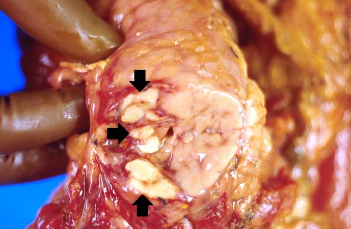
| 05:54, 21 August 2013 | IPLab13WT2.jpg (file) |  |
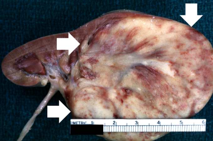
39 KB | This is a closer view of the kidney with Wilms' tumor (arrows). | 1 |
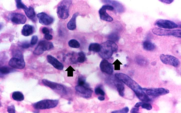
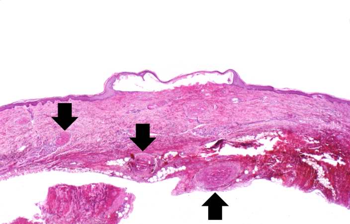
| 03:59, 21 August 2013 | IPLab9Actinomycosis1.jpg (file) |  |
39 KB | This is a low-power photomicrograph of the retroperitoneal abscess. At this magnification, multiple dark-staining foci can be appreciated. These foci are Actinomyces colonies (arrows). These colonies are known as "sulfur granules" because in gross spec... | 1 |
| 04:17, 21 August 2013 | IPLab10Blasto10.jpg (file) |  |
39 KB | This is a very high-power photomicrograph showing Blastomyces organisms stained with PAS. Note the budding organism (arrow) and the underlying pyogranulomatous inflammatory reaction in the background. | 1 |
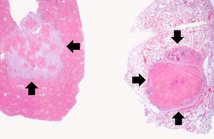
| 01:46, 21 August 2013 | IPLab7Metastatic3.jpg (file) |  |
39 KB | These are low-power photomicrographs of a section of liver (left) and lung (right) containing tumor nodules (arrows). | 1 |
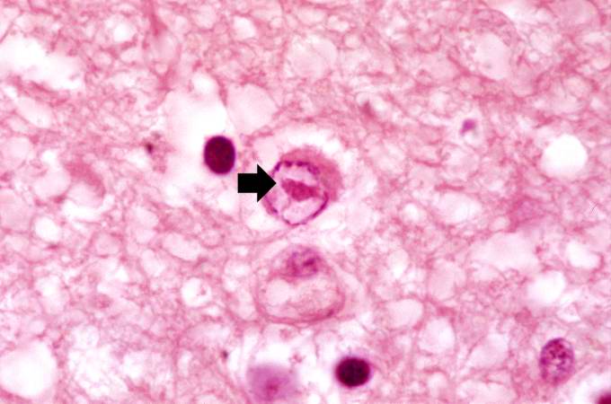
| 02:37, 21 August 2013 | IPLab8HSVEncephalitis10.jpg (file) |  |
40 KB | This is another high-power photomicrograph of a cell containing an intranuclear inclusion body (arrow). Note that the nuclear chromatin has been pushed to the outer edges of the nucleus. | 1 |
| 05:20, 21 August 2013 | IPLab12Acetaminophen7.jpg (file) |  |
40 KB | This low-power photomicrograph of the skin from this patient shows a blister and numerous thrombosed vessels (arrows) in the dermis. | 1 |
| 03:29, 19 August 2013 | IPLab3Sarcoidosis1.jpg (file) |  |
40 KB | This is a low-power photomicrograph of a lymph node. Note the rather pale-pink color of the tissue with dark-staining cells found in only a few scattered areas. These darker cells represent the original lymphocytes of this lymphoid organ. | 1 |
| 05:13, 21 August 2013 | IPLab12Alcoholic1.jpg (file) |  |
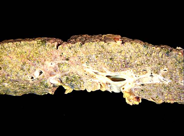
40 KB | This is a gross photograph of the liver from this patient. Note the nodular pattern and the areas of greenish discoloration as well as pale tan areas. | 1 |
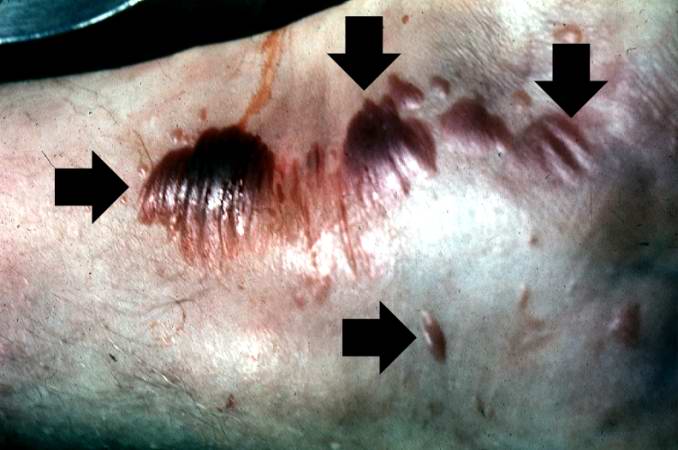
| 03:56, 21 August 2013 | IPLab9Clostridium2.jpg (file) |  |
40 KB | This gross photograph shows a close-up view of hemorrhagic blebs (arrows) on the skin. The blebs on the skin are accumulations of gas being discharged into the tissues from the Clostridium perfringens. This gas produces crepitance. | 1 |
| 04:19, 21 August 2013 | IPLab10Mucor4.jpg (file) |  |
40 KB | This is a higher-power photomicrograph of just the wall of the carotid artery. Note the ribbon-like clear structure with roughly parallel walls (non-septate hyphae) and right-angle branching (arrow). This is the Mucor organism. | 1 |
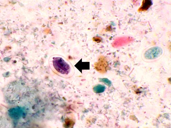
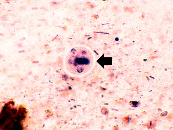
| 05:02, 21 August 2013 | IPLab11Ascariasis7.jpg (file) |  |
40 KB | This high-power photomicrograph of the fecal specimen from this patient shows a Giardia lamblia cyst (arrow). | 1 |
| 05:05, 21 August 2013 | IPLab11Schistosomiasis2.jpg (file) |  |
41 KB | This is high-power photomicrograph of the patient's fecal specimen containing another Schistosoma mansoni egg. | 1 |
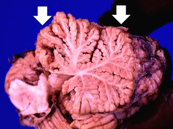
| 05:16, 21 August 2013 | IPLab12Alcoholic13.jpg (file) |  |
41 KB | This photograph of the cerebellum from this patient demonstrates the marked thinning of the anterior portion of the cerebellum (arrows). | 1 |
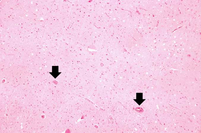
| 02:34, 21 August 2013 | IPLab8HSVEncephalitis3.jpg (file) |  |
41 KB | This is a low-power photomicrograph showing a section of brain with numerous perivascular hemorrhages (arrows) and some areas that appear hypercellular. | 1 |
| 05:23, 21 August 2013 | IPLab12RadiationFibrosis4.jpg (file) |  |
41 KB | This is a low-power photomicrograph of lung section. Note the thickening of the alveolar septa (arrows). | 1 |
| 05:01, 21 August 2013 | IPLab11Ascariasis5.jpg (file) |  |
41 KB | This high-power photomicrograph of the fecal specimen from this same patient shows an Entamoeba histolytica cyst (arrow). | 1 |
| 02:41, 21 August 2013 | IPLab8Rabies5.jpg (file) |  |
41 KB | This is a high-power photomicrograph of a neuron surrounded by inflammatory cells (lymphocytes and microglia). This neuron has two intracytoplasmic eosinophilic inclusion bodies (arrows). | 1 |
| 21:56, 20 August 2013 | IPLab6AcuteRejection2.jpg (file) |  |
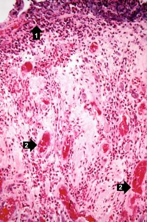
41 KB | This is a low-power photomicrograph of the kidney that was removed from this patient. Even at this low power you can appreciate the focal accumulations of cells within this section and the diffuse cellular infiltrate (blue dots) throughout the kidney p... | 1 |
| 05:15, 21 August 2013 | IPLab12Alcoholic9.jpg (file) |  |
41 KB | In this closer view of the distal esophagus the ruptured varix is indicated by the probe. Other varices and areas of submucosal hemorrhage can also be appreciated. | 1 |
| 16:07, 19 August 2013 | IPLab4PulmonaryCongestion4.jpg (file) |  |
42 KB | This is a low-power photomicrograph of lung from this case. The lung section has a pale-red color indicating proteinaceous material within the lung. | 1 |
| 16:35, 19 August 2013 | IPLab4Thrombosis2.jpg (file) |  |
42 KB | This is a low-power photomicrograph of thrombosed coronary artery. The thrombus (1) completely occludes the vessel. Note the layering of the thrombus. The fibrous cap is ruptured (arrow) and there is hemorrhage into the atherosclerotic plaque. Note the... | 1 |
| 01:17, 16 August 2013 | IPLab1FatNecrosis2.jpg (file) |  |
42 KB | 1 | |
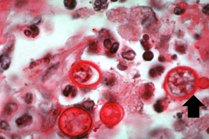
| 03:30, 19 August 2013 | IPLab3Sarcoidosis5.jpg (file) |  |
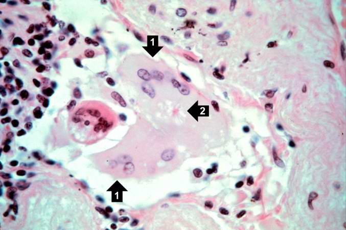
42 KB | This is a photomicrograph of a multinucleated giant cell (1). In the center of this foreign body-containing giant cell there is a small asteroid body (2). There is no functional significance to this asteroid body. | 1 |
| 05:13, 21 August 2013 | IPLab12Alcoholic6.jpg (file) |  |
42 KB | In this high-power photomicrograph of trichrome-stained liver, the bands of fibrous tissue surround the hepatocyte nodules. There is some degeneration and dropout of hepatocytes in this nodule. Also note the increased numbers of bile ducts in the triad... | 1 |