File list
This special page shows all uploaded files.
| Date | Name | Thumbnail | Size | Description | Versions |
|---|---|---|---|---|---|
| 18:07, 19 August 2013 | IPLab6RA7.jpg (file) |  |
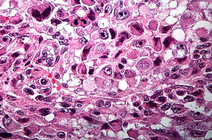
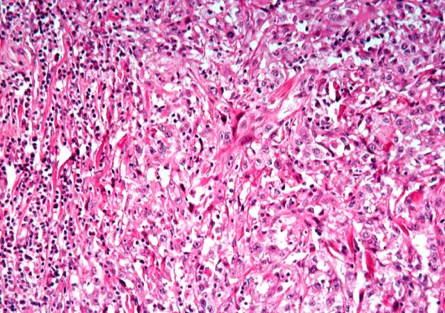
82 KB | This higher-power photomicrograph of the subcutaneous nodule again demonstrates the necrotic center and peripheral rim of macrophages, fibrocytes, and occasional lymphocytes. There are focal accumulations of hyaline material (fibrinoid material) within... | 1 |
| 16:12, 19 August 2013 | IPLab4PulmonaryCongestion7.jpg (file) |  |
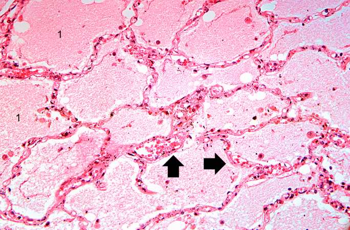
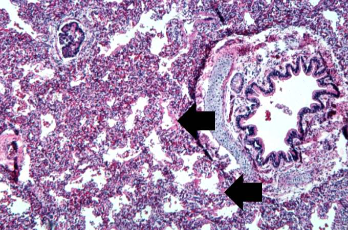
82 KB | This high-power photomicrograph illustrates the edema fluid within the alveoli (1) and the congestion (RBCs) in the alveolar capillaries (arrows). | 1 |
| 05:16, 21 August 2013 | IPLab12Alcoholic12.jpg (file) |  |
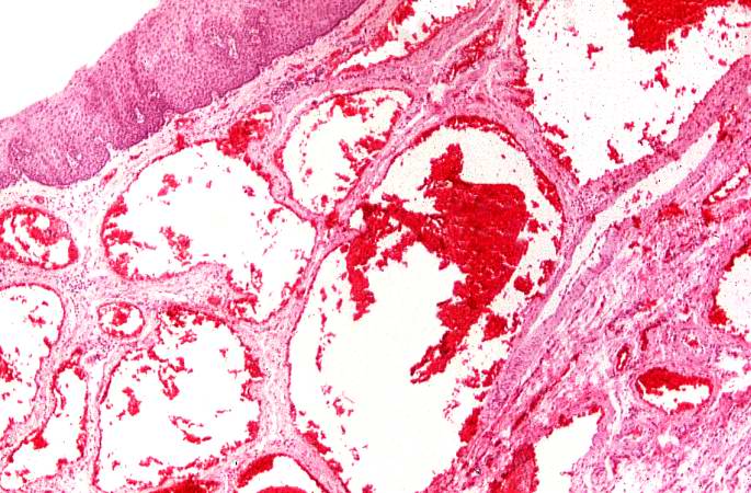
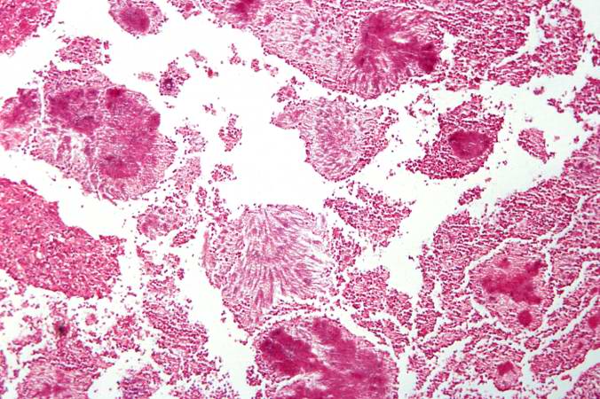
82 KB | This photomicrograph shows the dilated vessel just under the epithelium of the esophagus. | 1 |
| 02:44, 21 August 2013 | IPLab8Polio5.jpg (file) |  |
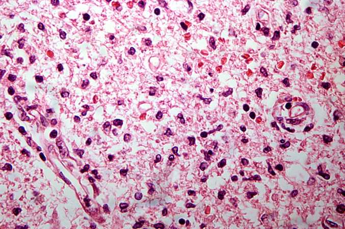
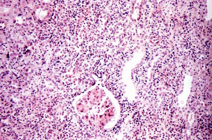
82 KB | This is another high-power photomicrograph of the anterior horn with inflammatory cell infiltrate and total loss of neurons. | 1 |
| 05:48, 21 August 2013 | IPLab13BiliaryAtresia3.jpg (file) |  |
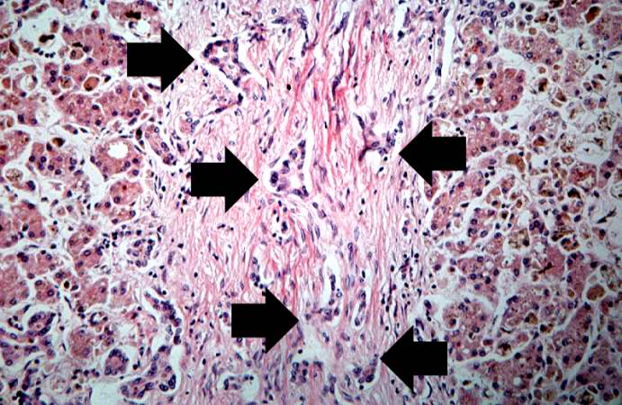
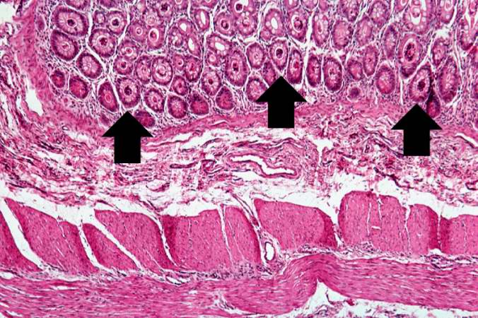
82 KB | This high-power photomicrograph of fibrotic portal region demonstrates proliferation of the bile ducts (arrows). | 1 |
| 02:47, 21 August 2013 | IPLab8HBV2.jpg (file) |  |
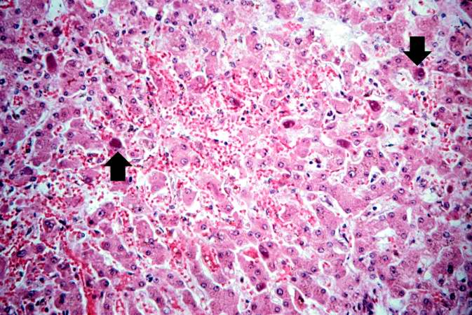
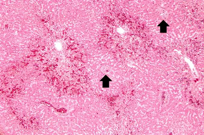
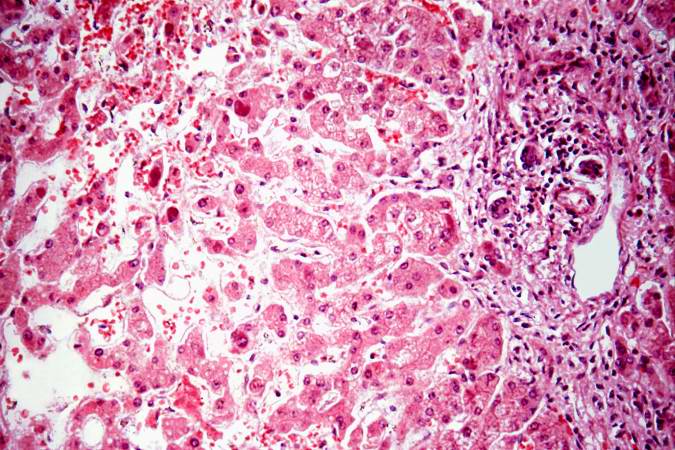
82 KB | This is a higher-power photomicrograph of liver from this case. Note the severe congestion (RBCs in sinusoids) and the presence of occasional hepatocytes with dark red/magenta-stained cytoplasm (arrows). | 1 |
| 01:56, 21 August 2013 | IPLab7Melanoma6.jpg (file) |  |
82 KB | This is a higher magnification showing the abundant extracellular melanin surrounding the tumor cells (brown pigment). | 1 |
| 02:15, 21 August 2013 | IPLab7Osteosarcoma10.jpg (file) |  |
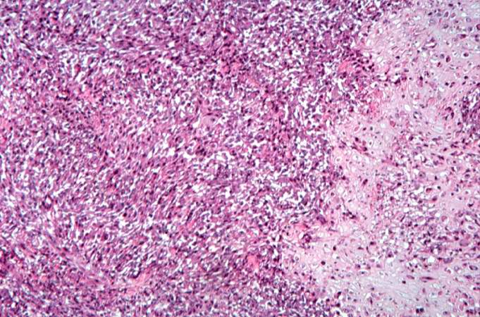
83 KB | This high-power photomicrograph demonstrates the growth pattern and the cell morphology. | 1 |
| 05:59, 21 August 2013 | IPLab13CF3.jpg (file) |  |
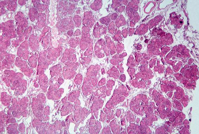
83 KB | This low-power photomicrograph of pancreas shows increased interstitial connective tissue resulting in accentuation of the lobular pattern. | 1 |
| 16:20, 19 August 2013 | IPLab4ChronicPassiveCongestion4.jpg (file) |  |
83 KB | This is a higher-power photomicrograph of liver demonstrating an accentuated lobular pattern with a dark red stain surrounding the central veins in the liver lobules (arrows). | 1 |
| 04:29, 19 August 2013 | IPLab3AcuteMyocardialInfarction3.jpg (file) |  |
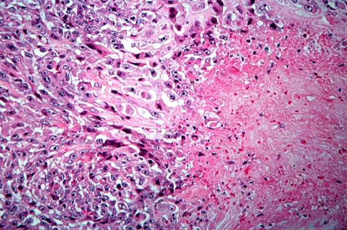
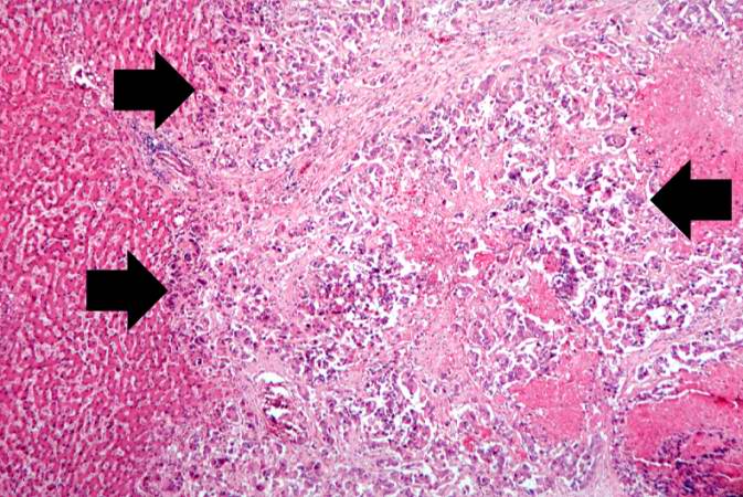
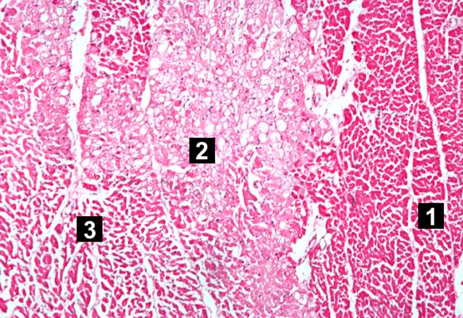
83 KB | This is a photomicrograph of the edge of the infarct with normal tissue on the left (1). The accumulation of inflammatory cells (2) is at the edge of the infarcted tissue (3). | 1 |
| 03:52, 21 August 2013 | IPLab9ARF3.jpg (file) |  |
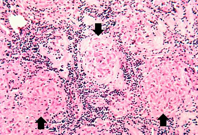
83 KB | This is a higher-power photomicrograph of myocardium showing cellular accumulations--Aschoff bodies (arrows)--within the interstitium of the myocardium. These are found especially around blood vessels. | 1 |
| 04:10, 21 August 2013 | IPLab10Crypto2.jpg (file) |  |
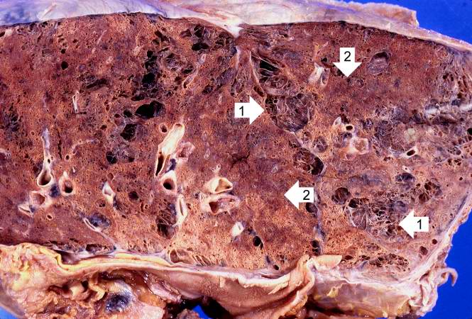
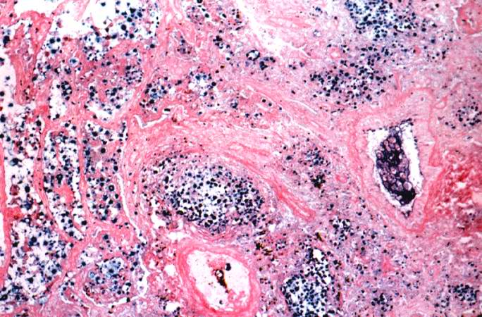
83 KB | This is a gross photomicrograph of this lung taken at autopsy. Note the areas of emphysema (1) and consolidation (2). | 1 |
| 02:16, 21 August 2013 | IPLab7Osteosarcoma13.jpg (file) |  |
83 KB | This is a high-power photomicrograph of the tumor demonstrating the anaplastic cell morphology. | 1 |
| 01:42, 21 August 2013 | IPLab7ColonCA9.jpg (file) |  |
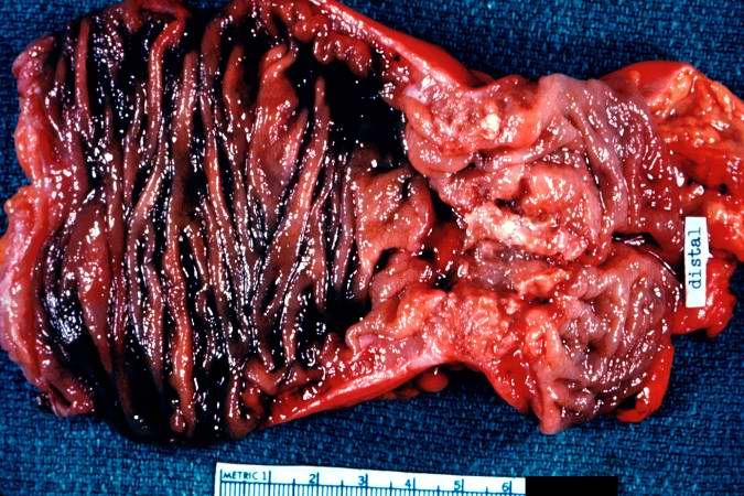
83 KB | This is a segment of distal colon from another case. Note the annular tumor that severely compromises the lumen of the colon. There is dilation of the colon proximal to the tumor. | 1 |
| 01:18, 16 August 2013 | IPLab1FatNecrosis6.jpg (file) |  |
83 KB | 1 | |
| 21:57, 20 August 2013 | IPLab6AcuteRejection5.jpg (file) |  |
83 KB | This is a higher-power photomicrograph demonstrating the cellular infiltrate within the interstitium and around the small blood vessel in the center of the image. | 1 |
| 03:56, 21 August 2013 | IPLab9Clostridium4.jpg (file) |  |
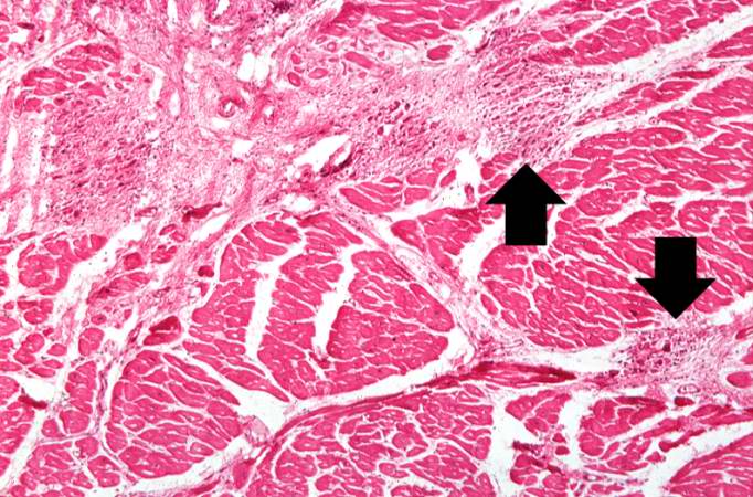
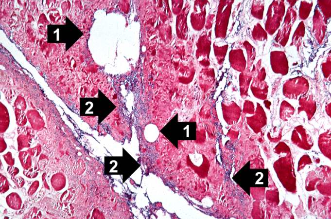
83 KB | This is a high-power photomicrograph of skeletal muscle. The muscle cells are hypereosinophilic and most do not contain nuclei, indicating that these cells are dead or dying. The round clear spaces (1) in this tissue correspond to gas accumulations pri... | 1 |
| 16:20, 19 August 2013 | IPLab4ChronicPassiveCongestion5.jpg (file) |  |
83 KB | This higher-power photomicrograph of the liver lobules shows congestion and red blood cell accumulation in the sinusoidal spaces around the central vein. Note that around the portal triads (arrows) the liver cells are quite normal and there is no evide... | 1 |
| 03:30, 19 August 2013 | IPLab3Sarcoidosis3.jpg (file) |  |
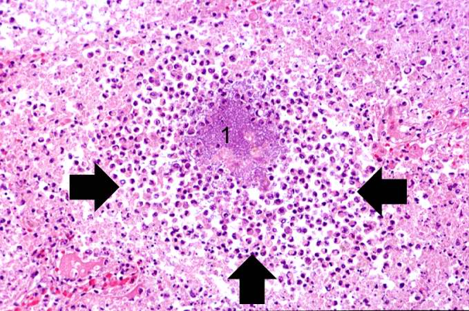
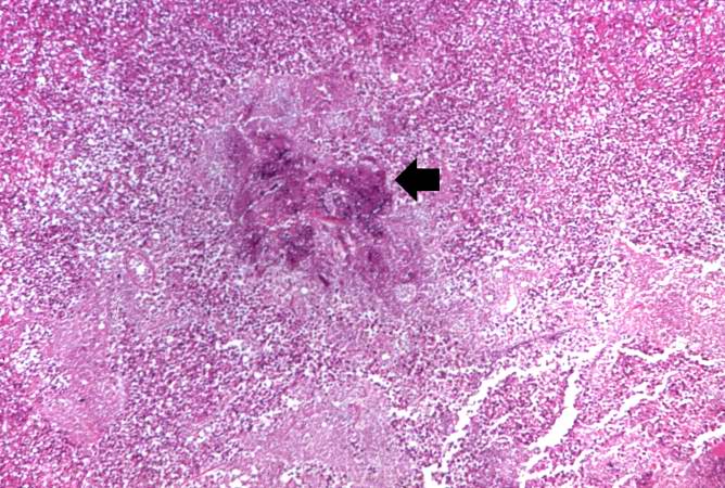
84 KB | This is a photomicrograph of the small nodules (arrows) seen in the previous image. Close examination reveals that they are composed of large macrophages (epithelioid macrophages). These small granulomas form multiple series of reaction centers through... | 1 |
| 21:49, 20 August 2013 | IPLab6ChronicRejection4.jpg (file) |  |
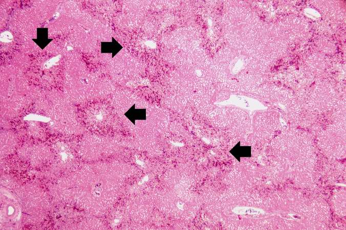
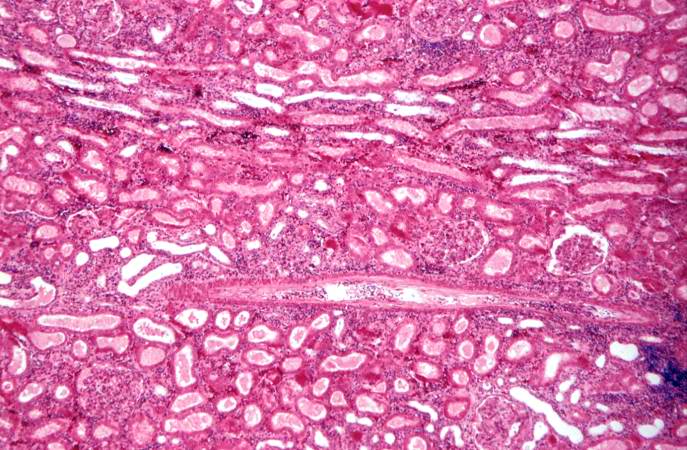
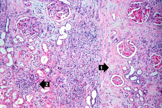
84 KB | This is another area of renal cortex similar to the previous image. Note the fibrosis (1) and loss of renal tubules throughout this section. Also note the focus of inflammatory cells (2) indicating that despite the chromic nature of this lesion, there ... | 1 |
| 05:55, 21 August 2013 | IPLab13WT6.jpg (file) |  |
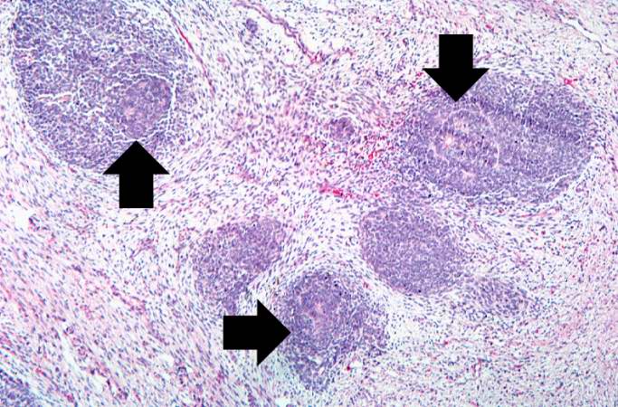
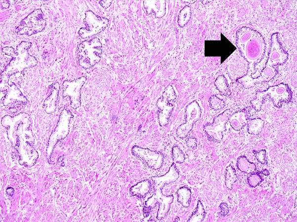
84 KB | This medium-power photomicrograph of tumor shows again the two cell types making up this neoplasm. There are regions within the blastema where the cells form glands or "tubules" (arrows). | 1 |
| 04:12, 21 August 2013 | IPLab10Crypto10.jpg (file) |  |
84 KB | This is a low-power photomicrograph of lung section stained with Alcian blue, which stains the acidic glycosaminoglycans making up the coat of the cryptococcal organism. | 1 |
| 05:51, 21 August 2013 | IPLab13Hyaline6.jpg (file) |  |
85 KB | This is a medium-power photomicrograph showing a large bronchus with cartilage. Interstitial congestion with numerous red cells is apparent. Even at this magnification hyaline membranes (arrows) can be seen lining the alveoli. | 1 |
| 01:32, 21 August 2013 | IPLab7LipSCC6.jpg (file) |  |
85 KB | This is a high power photomicrograph of the well-differentiated squamous cell carcinoma. Note the intracytoplasmic keratinization which gives the cells a glassy appearance. The focal accumulations of keratinized cells are called keratin pearls (arrows). | 1 |
| 21:36, 20 August 2013 | IPLab6Amyloid10.jpg (file) |  |
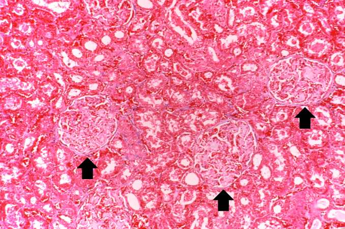
85 KB | This photomicrograph of kidney demonstrates the amyloid deposits (arrows) within glomeruli. | 1 |
| 02:48, 21 August 2013 | IPLab8HBV3.jpg (file) |  |
85 KB | This is a higher-power photomicrograph of the periportal region exhibiting some inflammation and bile duct hyperplasia. There is also congestion and some loss of hepatocytes with disruption of the hepatic cords. | 1 |
| 06:00, 21 August 2013 | IPLab13CF4.jpg (file) |  |
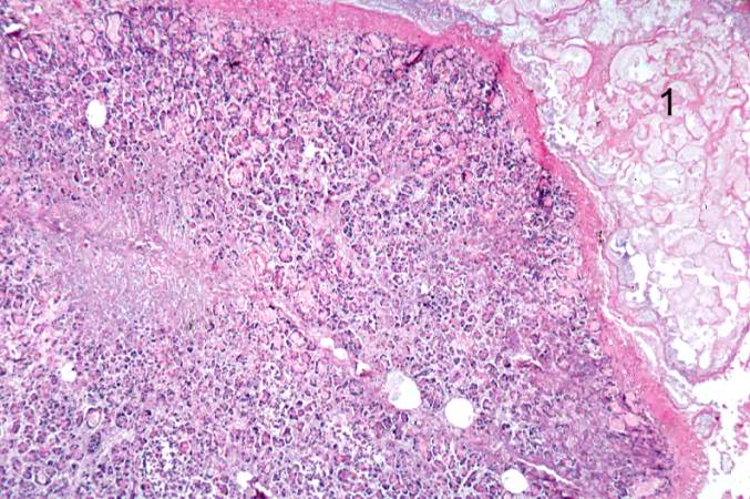
85 KB | This higher-power photomicrograph of the pancreas shows interstitial tissue and the presence of small cystic spaces (1) within the acinar lobules. These spaces are filled with an eosinophilic proteinaceous material. The islets of Langerhans (2) are una... | 1 |
| 21:34, 20 August 2013 | IPLab6Amyloid5.jpg (file) |  |
85 KB | This is a higher-power photomicrograph showing the amyloid deposits (1) between hepatocytes (2). | 1 |
| 01:46, 21 August 2013 | IPLab7Metastatic5.jpg (file) |  |
85 KB | This is a higher-power photomicrograph showing how the tumor cells (arrows) have infiltrated into the liver parenchyma. | 1 |
| 02:16, 21 August 2013 | IPLab7Osteosarcoma15.jpg (file) |  |
85 KB | This is a high-power photomicrograph of the tumor demonstrating the anaplastic cell morphology and multiple mitotic figures (arrows). | 1 |
| 16:15, 15 August 2013 | IPLab1LungAbscess8.jpg (file) |  |
85 KB | 1 | |
| 01:33, 21 August 2013 | IPLab7LipSCC7.jpg (file) |  |
85 KB | This is a high power photomicrograph of a poorly-differentiated area of tumor. Note the spindle-shaped cells and the irregular pattern of growth. | 1 |
| 13:50, 15 August 2013 | IPLab1MyocardialInfarction4.jpg (file) |  |
85 KB | 1 | |
| 21:50, 20 August 2013 | IPLab6ChronicRejection10.jpg (file) |  |
86 KB | This is a high-power photomicrograph of a kidney from another case of chronic transplant rejection. In this case there is extensive damage to the kidney due to the chronic rejection (loss of tubules and glomerular lesions). In addition, this kidney was... | 1 |
| 03:59, 21 August 2013 | IPLab9Actinomycosis3.jpg (file) |  |
86 KB | This is a higher-power photomicrograph of actinomycotic colonies in the abscess. | 1 |
| 01:37, 21 August 2013 | IPLab7EsophSCC4.jpg (file) |  |
86 KB | This is a higher-power photomicrograph showing invasive squamous cell carcinoma. Tongues and islands of tumor cells exhibit areas of central necrosis (arrow). | 1 |
| 06:01, 21 August 2013 | IPLab13CF12.jpg (file) |  |
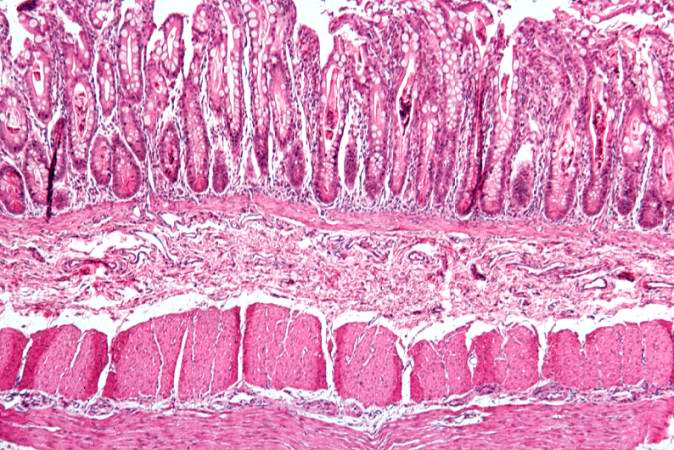
86 KB | This is a low-power photomicrograph from another section of the intestine. Saggital sections of the intestinal crypts show the crypts along their full length, extending to the mucosal surface. | 1 |
| 03:23, 19 August 2013 | IPLab3Bronchopneumonia7.jpg (file) |  |
86 KB | This higher-power photomicrograph shows a central portion of an abscess. Note the absence of any parenchymal lung tissue in this section due to extensive neutrophilic infiltration with liquefaction necrosis of the parenchymal tissue. Masses of leukocyt... | 1 |
| 02:15, 21 August 2013 | IPLab7Osteosarcoma9.jpg (file) |  |
86 KB | This high-power photomicrograph demonstrates the cellular growth pattern. Note that the cells are fusiform and they grow in sheets. | 1 |
| 03:40, 16 August 2013 | IPLab1Prostate1.jpg (file) |  |
86 KB | 1 | |
| 05:48, 21 August 2013 | IPLab13BiliaryAtresia5.jpg (file) |  |
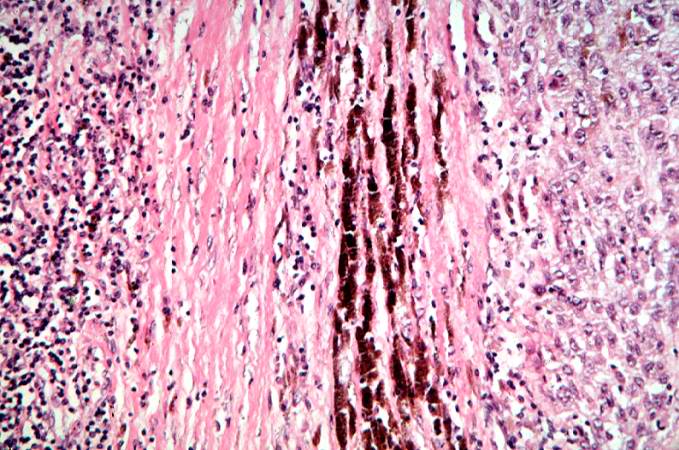
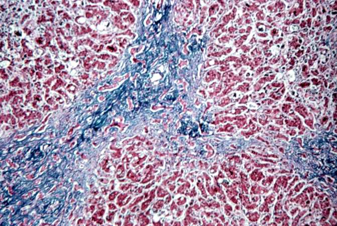
87 KB | This is a medium-power photomicrograph of liver section stained with a trichrome stain to demonstrate the portal fibrosis. The fibrous connective tissue (collagen) stains blue. | 1 |
| 06:01, 21 August 2013 | IPLab13CF9.jpg (file) |  |
87 KB | A higher-power photomicrograph shows the bottom of the intestinal crypts and the other normal layers of the intestine. Even at this magnification, accumulations of eosinophilic debris can be seen in many of the intestinal crypts (arrows). | 1 |
| 01:41, 21 August 2013 | IPLab7ColonCA5.jpg (file) |  |
88 KB | This is a high-power photomicrograph of tumor cells invading the underlying muscularis. | 1 |
| 21:58, 20 August 2013 | IPLab6AcuteRejection8.jpg (file) |  |
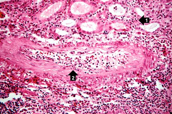
88 KB | This high-power photomicrograph demonstrates the cellular infiltrate within the interstitium (1) and in the wall of the blood vessel (2). | 1 |
| 01:23, 21 August 2013 | IPLab7Adenoma5.jpg (file) |  |
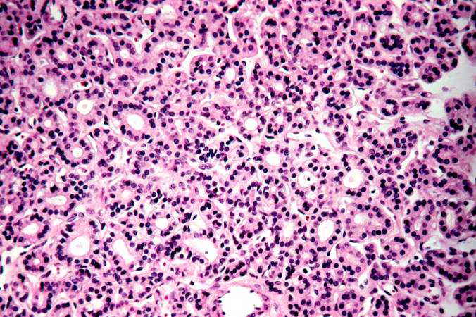
88 KB | This is a photomicrograph of an adenoma. Note that the follicular architecture is well developed and more or less uniform throughout this section. | 1 |
| 02:12, 19 August 2013 | IPLab3AcuteAppendicitis5.jpg (file) |  |
88 KB | This photomicrograph of the mucosal surface shows a small area with normal mucosal epithelium (arrow). This area is surrounded by areas of ulceration with an inflammatory infiltrate of lymphocytes and neutrophils. | 1 |
| 04:30, 19 August 2013 | IPLab3AcuteMyocardialInfarction7.jpg (file) |  |
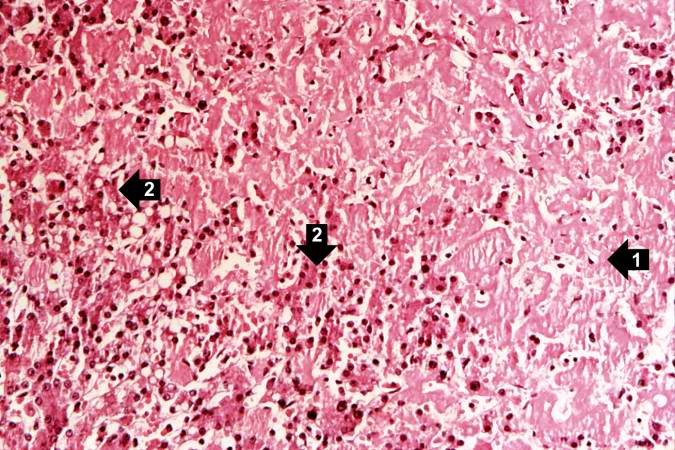
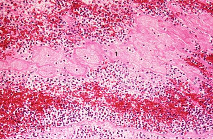
88 KB | This is a photomicrograph of the lines of Zahn. Pale areas (1) represent platelets with some fibrin and the darker lines (2) represent RBCs and leukocytes enmeshed in fibrin strands. | 1 |
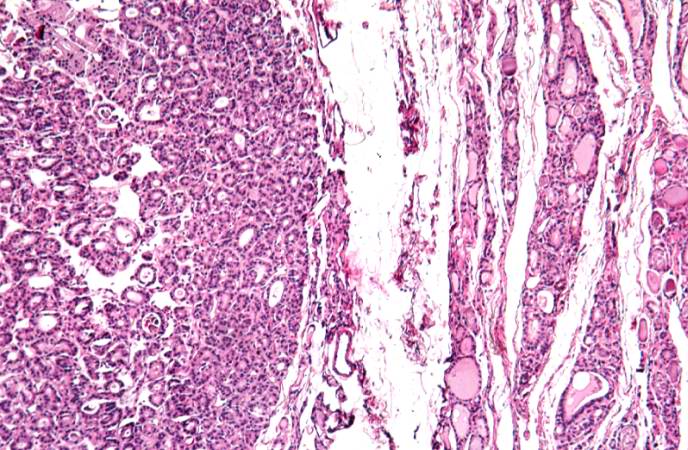
| 01:22, 21 August 2013 | IPLab7Adenoma3.jpg (file) |  |
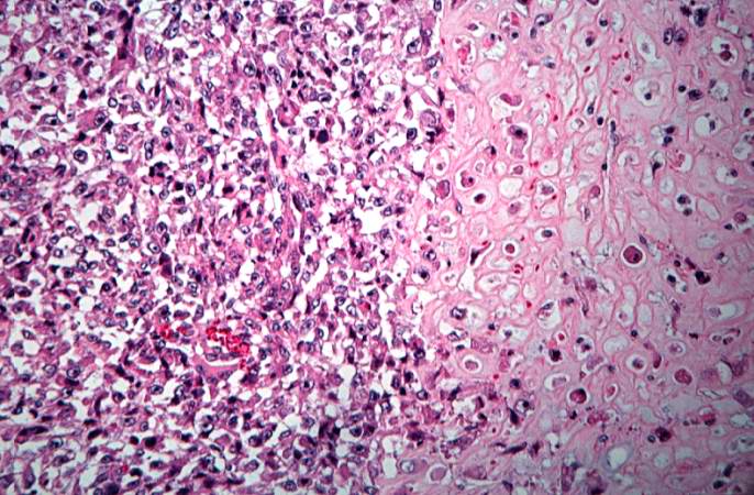
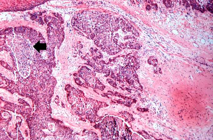
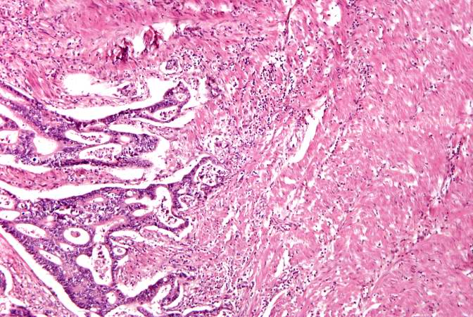
89 KB | This is another higher-power photomicrograph of the adenoma (left) and the adjacent thyroid tissue (right). Note the compression of the adjacent normal thyroid and the difference in morphology between the adenoma and the thyroid. | 1 |
| 03:47, 19 August 2013 | IPLab3ForeignBodyGranuloma3.jpg (file) |  |
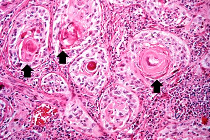
89 KB | A higher-power photomicrograph of these granulomas reveals that they surround blood vessels (note the red blood cells within the lumen) (arrow). | 1 |