Difference between revisions of "IPLab:Lab 7:Carcinoid"
Seung Park (talk | contribs) |
(→Images) |
||
| (11 intermediate revisions by 2 users not shown) | |||
| Line 1: | Line 1: | ||
== Clinical Summary == | == Clinical Summary == | ||
| − | This 58-year-old male experienced increasing diarrhea | + | This 58-year-old male experienced increasing diarrhea over the 4 months prior to admission. During this period he experienced a weight loss of 40 pounds. Imaging demonstrated a lesion a the ileocecal valve and a laparotomy was performed. |
| − | + | The operative specimen consisted of 12 cm of distal ileum, appendix, cecum and colon. On opening the bowel there was a 4.5 x 3 x 3-cm elliptical submucosal mass at the ileocecal valve that had produced partial obstruction. Several small (2 mm) submucosal masses were found in the cecum nearby. On cut section each lesion was found to be firm, gray-tan and homogeneous involving the muscular wall of the bowel and adjacent mesentery. | |
| − | The operative specimen consisted of 12 cm of distal ileum, appendix, cecum and | ||
== Images == | == Images == | ||
<gallery heights="250px" widths="250px"> | <gallery heights="250px" widths="250px"> | ||
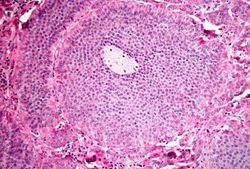
File:IPLab7Carcinoid1.jpg|This is a low-power photomicrograph of the surgical specimen showing basophilic and eosinophilic areas delimiting areas of tumor infiltration. | File:IPLab7Carcinoid1.jpg|This is a low-power photomicrograph of the surgical specimen showing basophilic and eosinophilic areas delimiting areas of tumor infiltration. | ||
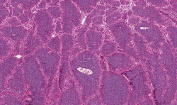
| − | File: | + | File:IPLab7Carcinoid1x.jpg|This is a higher-power photomicrograph of the surgical specimen showing the mucosa is normal and the tumor cells are in the submucosa. |
| − | File: | + | File:IPLab7Carcinoid3b.jpg|This is a higher-power photomicrograph of the surgical specimen showing nests of tumor cells. |
| + | File:IPLab7Carcinoid4b.jpg|This is a high-power photomicrograph of the surgical specimen showing the tumor's growth pattern--cells form discrete islands, trabeculae, and glands. | ||
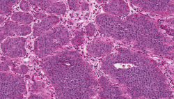
File:IPLab7Carcinoid4.jpg|This is a high-power photomicrograph of the surgical specimen showing the cellular morphology. The tumor cells are monotonously similar with scant, pink, granular cytoplasm and a round-to-oval stippled nucleus. As in most carcinoid tumors, there is minimal variation in cell and nuclear size, and mitoses are infrequent or absent. | File:IPLab7Carcinoid4.jpg|This is a high-power photomicrograph of the surgical specimen showing the cellular morphology. The tumor cells are monotonously similar with scant, pink, granular cytoplasm and a round-to-oval stippled nucleus. As in most carcinoid tumors, there is minimal variation in cell and nuclear size, and mitoses are infrequent or absent. | ||
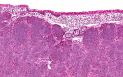
| − | File: | + | File:IPLab7Carcinoid8x.jpg|This low-power photomicrograph shows how the tumor cells have invaded into the muscularis of the bowel (arrows). |
| − | |||
| − | |||
| − | |||
| − | |||
| − | |||
| − | |||
</gallery> | </gallery> | ||
| + | |||
| + | == Virtual Microscopy == | ||
| + | <peir-vm>IPLab7Carcinoid</peir-vm> | ||
== Study Questions == | == Study Questions == | ||
| Line 24: | Line 21: | ||
* <spoiler text="What is 'carcinoid syndrome' and how common is it?">Carcinoid syndrome occurs in about 1% of all patients with carcinoids and in 20% of those with widespread metastases. Uncertainties remain about the precise origin of the carcinoid syndrome, but most manifestations are thought to arise from excess elaboration of serotonin. The clinical syndrome can include cutaneous flushes and apparent cyanosis; intestinal hypermotility (diarrhea, cramps, nausea, vomiting); asthmatic bronchoconstrictive attacks; hepatomegaly (due to metastases); systemic fibrosis (cardiac, aortic, retroperinoteal. pelvic).</spoiler> | * <spoiler text="What is 'carcinoid syndrome' and how common is it?">Carcinoid syndrome occurs in about 1% of all patients with carcinoids and in 20% of those with widespread metastases. Uncertainties remain about the precise origin of the carcinoid syndrome, but most manifestations are thought to arise from excess elaboration of serotonin. The clinical syndrome can include cutaneous flushes and apparent cyanosis; intestinal hypermotility (diarrhea, cramps, nausea, vomiting); asthmatic bronchoconstrictive attacks; hepatomegaly (due to metastases); systemic fibrosis (cardiac, aortic, retroperinoteal. pelvic).</spoiler> | ||
* <spoiler text="What is the prognosis for patients with carcinoid tumors?">The overall 5-year survival rate for carcinoid tumors (excluding appendiceal) is approximately 90%. Even with small bowel tumors and hepatic metastases, there is a 50% 5-year survival. Widespread disease, however, usually causes death.</spoiler> | * <spoiler text="What is the prognosis for patients with carcinoid tumors?">The overall 5-year survival rate for carcinoid tumors (excluding appendiceal) is approximately 90%. Even with small bowel tumors and hepatic metastases, there is a 50% 5-year survival. Widespread disease, however, usually causes death.</spoiler> | ||
| + | |||
| + | == Additional Resources == | ||
| + | === Reference === | ||
| + | * [http://emedicine.medscape.com/article/282515-overview eMedicine Medical Library: Malignant Carcinoid Syndrome] | ||
| + | * [http://www.merckmanuals.com/professional/endocrine_and_metabolic_disorders/carcinoid_tumors/overview_of_carcinoid_tumors.html Merck Manual: Overview of Carcinoid Tumors] | ||
| + | * [http://www.merckmanuals.com/professional/endocrine_and_metabolic_disorders/carcinoid_tumors/carcinoid_syndrome.html Merck Manual: Carcinoid Syndrome] | ||
| + | |||
| + | === Journal Articles === | ||
| + | * Shebani KO, Souba WW, Finkelstein DM, Stark PC, Elgadi KM, Tanabe KK, Ott MJ. [http://www.ncbi.nlm.nih.gov/pubmed/10363895 Prognosis and survival in patients with gastrointestinal tract carcinoid tumors]. ''Ann Surg'' 1999 Jun;229(6):815-21; discussion 822-3. | ||
| + | |||
| + | === Images === | ||
| + | * [{{SERVER}}/library/index.php?/tags/1592-carcinoid PEIR Digital Library: Carcinoid Images] | ||
| + | * [http://library.med.utah.edu/WebPath/GIHTML/GIIDX.html#5 WebPath: Small Intestine] | ||
{{IPLab 7}} | {{IPLab 7}} | ||
[[Category: IPLab:Lab 7]] | [[Category: IPLab:Lab 7]] | ||
Latest revision as of 03:39, 9 July 2020
Contents
Clinical Summary[edit]
This 58-year-old male experienced increasing diarrhea over the 4 months prior to admission. During this period he experienced a weight loss of 40 pounds. Imaging demonstrated a lesion a the ileocecal valve and a laparotomy was performed.
The operative specimen consisted of 12 cm of distal ileum, appendix, cecum and colon. On opening the bowel there was a 4.5 x 3 x 3-cm elliptical submucosal mass at the ileocecal valve that had produced partial obstruction. Several small (2 mm) submucosal masses were found in the cecum nearby. On cut section each lesion was found to be firm, gray-tan and homogeneous involving the muscular wall of the bowel and adjacent mesentery.
Images[edit]
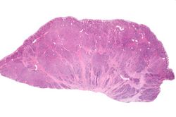
This is a low-power photomicrograph of the surgical specimen showing basophilic and eosinophilic areas delimiting areas of tumor infiltration.
This is a higher-power photomicrograph of the surgical specimen showing the mucosa is normal and the tumor cells are in the submucosa.
This is a higher-power photomicrograph of the surgical specimen showing nests of tumor cells.
This is a high-power photomicrograph of the surgical specimen showing the tumor's growth pattern--cells form discrete islands, trabeculae, and glands.
This is a high-power photomicrograph of the surgical specimen showing the cellular morphology. The tumor cells are monotonously similar with scant, pink, granular cytoplasm and a round-to-oval stippled nucleus. As in most carcinoid tumors, there is minimal variation in cell and nuclear size, and mitoses are infrequent or absent.
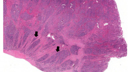
This low-power photomicrograph shows how the tumor cells have invaded into the muscularis of the bowel (arrows).


Virtual Microscopy[edit]
Study Questions[edit]
Additional Resources[edit]
Reference[edit]
- eMedicine Medical Library: Malignant Carcinoid Syndrome
- Merck Manual: Overview of Carcinoid Tumors
- Merck Manual: Carcinoid Syndrome
Journal Articles[edit]
- Shebani KO, Souba WW, Finkelstein DM, Stark PC, Elgadi KM, Tanabe KK, Ott MJ. Prognosis and survival in patients with gastrointestinal tract carcinoid tumors. Ann Surg 1999 Jun;229(6):815-21; discussion 822-3.
Images[edit]
Cyanosis is a bluish discoloration of the skin and mucous membranes resulting from increased concentrations of reduced hemoglobin in the blood. Cyanosis occurs when the blood oxygen saturation falls below 85%.
The normal fibrinogen level is 184 to 412 mg/dL.