Difference between revisions of "IPLab:Lab 7:Carcinoid"
Seung Park (talk | contribs) (→Reference) |
Seung Park (talk | contribs) (→Journal Articles) |
||
| Line 32: | Line 32: | ||
=== Journal Articles === | === Journal Articles === | ||
| − | + | * Shebani KO, Souba WW, Finkelstein DM, Stark PC, Elgadi KM, Tanabe KK, Ott MJ. [http://www.ncbi.nlm.nih.gov/pubmed/10363895 Prognosis and survival in patients with gastrointestinal tract carcinoid tumors]. ''Ann Surg'' 1999 Jun;229(6):815-21; discussion 822-3. | |
=== Images === | === Images === | ||
Revision as of 20:01, 23 August 2013
Contents
Clinical Summary[edit]
This 58-year-old male experienced increasing diarrhea (up to 10-12 yellow watery stools per day) over the year prior to admission. During this period he experienced a weight loss of 40 pounds. A laparotomy was performed after a careful workup.
Autopsy Findings[edit]
The operative specimen consisted of 12 cm of distal ileum, appendix, cecum and 50 cm of colon. The ileum was mildly dilated and its wall hypertrophied. On opening the bowel there was a 4.5 x 3 x 3-cm elliptical submucosal mass at the ileocecal valve; this had produced partial obstruction. Several small (2 mm) submucosal masses were found in the cecum nearby. On cut section each lesion was found to be firm, gray-tan and homogeneous involving the muscular wall of the bowel and adjacent mesentery. The appendix and colon showed no lesions.
Images[edit]
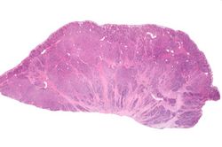
This is a low-power photomicrograph of the surgical specimen showing basophilic and eosinophilic areas delimiting areas of tumor infiltration.
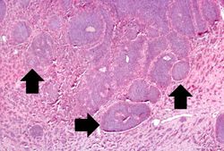
This is a higher-power photomicrograph of the surgical specimen showing nests of tumor cells (arrows).
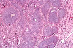
This is a high-power photomicrograph of the surgical specimen showing the tumor's growth pattern--cells form discrete islands, trabeculae, and glands.
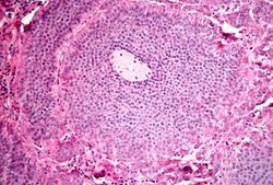
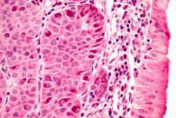
This is a high-power photomicrograph of the surgical specimen showing the cellular morphology. The tumor cells are monotonously similar with scant, pink, granular cytoplasm and a round-to-oval stippled nucleus. As in most carcinoid tumors, there is minimal variation in cell and nuclear size, and mitoses are infrequent or absent.
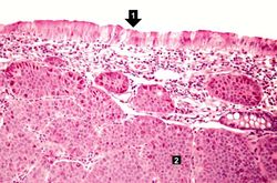
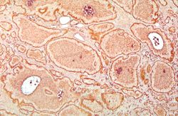
This is a low-power photomicrograph of one of the subcutaneous masses in the cecum. Note that the mucosa (1) is virtually normal and the tumor cells are in the submucosa (2).
This is a higher-power photomicrograph of the previous section showing the intact mucosa (right) and the submucosal carcinoid tumor.
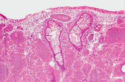
This is a low-power photomicrograph of another one of the subcutaneous masses in the cecum. The mucosa is normal and the tumor cells are in the submucosa.
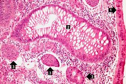
This is a higher-power photomicrograph of the previous section showing intact mucosa (1), a gland (2), and the submucosal carcinoid tumor cells (3).
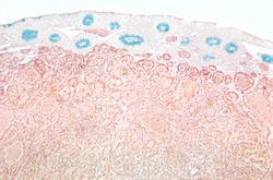
This is a low-power photomicrograph of a section of cecum containing tumor stained to demonstrate the secretory granules in these tumor cells (brown-colored stain). The blue color is the mucin in the glands just under the mucosal surface.
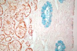
This is a higher-power view of the previous section stained with a silver stain to delineate carcinoid tumor cells (brown) and a mucin stain (blue) to stain the glands.
This is a high-power view of the same section stained with a silver stain to delineate carcinoid tumor cells (brown).


Study Questions[edit]
Additional Resources[edit]
Reference[edit]
- eMedicine Medical Library: Malignant Carcinoid Syndrome
- Merck Manual: Overview of Carcinoid Tumors
- Merck Manual: Carcinoid Syndrome
Journal Articles[edit]
- Shebani KO, Souba WW, Finkelstein DM, Stark PC, Elgadi KM, Tanabe KK, Ott MJ. Prognosis and survival in patients with gastrointestinal tract carcinoid tumors. Ann Surg 1999 Jun;229(6):815-21; discussion 822-3.
Images[edit]
Related IPLab Cases[edit]
Cyanosis is a bluish discoloration of the skin and mucous membranes resulting from increased concentrations of reduced hemoglobin in the blood. Cyanosis occurs when the blood oxygen saturation falls below 85%.
The normal fibrinogen level is 184 to 412 mg/dL.