Difference between revisions of "IPLab:Lab 7:Adenocarcinoma"
Seung Park (talk | contribs) |
(→Autopsy Findings) |
||
| (8 intermediate revisions by 2 users not shown) | |||
| Line 1: | Line 1: | ||
== Clinical Summary == | == Clinical Summary == | ||
| − | Approximately four months prior to admission, this 68-year-old male began having "sharp, shooting pains" in the lower abdomen. | + | Approximately four months prior to admission, this 68-year-old male began having "sharp, shooting pains" in the lower abdomen. On admission a CT scan showed a mass in the transverse colon. The patient refused to undergo a laparotomy and declined further treatment. Six months later he returned with severe abdominal pain and a colectomy was performed. |
| − | + | The segment of colon contained numerous polyps and an annular tumor which was 6.7 cm in diameter. Endoscopic examination of the ascending colon revealed two more polyps which were removed. | |
| − | The segment of colon contained numerous polyps and an annular tumor which was 6.7 cm in diameter. | ||
== Images == | == Images == | ||
| Line 17: | Line 16: | ||
File:IPLab7ColonCA9.jpg|This is a segment of distal colon from another case. Note the annular tumor that severely compromises the lumen of the colon. There is dilation of the colon proximal to the tumor. | File:IPLab7ColonCA9.jpg|This is a segment of distal colon from another case. Note the annular tumor that severely compromises the lumen of the colon. There is dilation of the colon proximal to the tumor. | ||
</gallery> | </gallery> | ||
| + | |||
| + | == Virtual Microscopy == | ||
| + | <peir-vm>IPLab7ColonCA</peir-vm> | ||
| + | |||
| + | == Study Questions == | ||
| + | * <spoiler text="What age group usually presents with colon cancer and what factors may predispose to early onset?">The peak incidence for colorectal carcinoma is age 60 to 70 years; fewer than 20% of cases occur under 50 years of age. When colorectal carcinoma is found in a young person, pre-existing ulcerative colitis or one of the polyposis syndromes must be suspected.</spoiler> | ||
| + | * <spoiler text="What is the usual presenting signs or symptoms of a patient with colon cancer?">The clinical presentation depends upon the site of the tumor. Colorectal cancers remain asymptomatic for years. Cecal and right colonic cancers most often lead to fatigue, weakness, and iron-deficiency anemia. These fungating lesions bleed easily and may be discovered at an early stage, provided that the colon is examined thoroughly radiographically and during colonoscopy. Left-sided lesions produce occult bleeding, changes in bowel habit, or crampy left lower quadrant discomfort. In theory, the chance for early discovery and successful removal should be greater with lesions on the left side because these patients usually have prominent disturbances in bowel function, such as melena, diarrhea, and constipation.</spoiler> | ||
| + | * <spoiler text="How does the morphology of colon cancer differ depending upon location?">Almost all colorectal carcinomas begin as in situ lesions within adenomatous polyps; however, they evolve into different morphologic patterns. Tumors in the proximal colon tend to grow as polypoid, fungating masses that extend along one wall of the capacious cecum and ascending colon. Obstruction is uncommon. When carcinomas in the distal colon are discovered, they tend to be annular, encircling lesions that produce so-called napkin-ring constrictions of the bowel. The margins of the napkin ring are classically heaped up, beaded, and firm, and the midregion is ulcerated. The lumen is markedly narrowed, and the proximal bowel may be distended.</spoiler> | ||
| + | * <spoiler text="What predisposing factors lead to colon cancer?">Dietary factors that may predispose to cancer are | ||
| + | # a low fiber diet, | ||
| + | # high intake of refined carbohydrates, | ||
| + | # high fat diet, and | ||
| + | # decreased intake of protective vitamins. | ||
| + | It is thought that reduced fiber content leads to decreased stool bulk, increased fecal transit time in the bowel, and an altered bacterial flora of the intestine. Potentially toxic oxidative byproducts of carbohydrate degradation by bacteria are therefore present in higher concentrations in the small stools and are held in contact with the colonic mucosa for longer periods of time. More recent epidemiologic data have raised some doubt about the importance of fiber in the diet but most nutritionalists still recommend high fiber diet to help prevent colon cancer. Moreover, high fat intake enhances the synthesis of cholesterol and bile acids by the liver, which in turn may be converted into potential carcinogens by intestinal bacteria. Refined diets also contain less vitamins A, C, and E, which may act as oxygen radical scavengers.</spoiler> | ||
| + | |||
| + | == Additional Resources == | ||
| + | === Reference === | ||
| + | * [http://emedicine.medscape.com/article/367061-overview eMedicine Medical Library: Imaging in Adenocarcinoma of the Colon] | ||
| + | * [http://emedicine.medscape.com/article/277496-overview eMedicine Medical Library: Colon Adenocarcinoma] | ||
| + | * [http://www.merckmanuals.com/professional/gastrointestinal_disorders/tumors_of_the_gi_tract/colorectal_cancer.html Merck Manual: Colorectal Cancer] | ||
| + | |||
| + | === Journal Articles === | ||
| + | * Yuen ST, Wong MP, Chung LP, Chan SY, Cheung N, Ho J, Leung SY. [http://www.ncbi.nlm.nih.gov/pubmed/9543668 Up-regulation of lysozyme production in colonic adenomas and adenocarcinomas]. ''Histopathology'' 1998 Feb;32(2):126-32. | ||
| + | * Strum WB. [http://www.nejm.org/doi/full/10.1056/NEJMra1513581 Colorectal Adenomas]. ''NEJM'' 2016 March 17 374:1065-1075. | ||
| + | |||
| + | === Images === | ||
| + | * [{{SERVER}}/library/index.php?/tags/66-colon/112-carcinoma PEIR Digital Library: Colon Carcinoma Images] | ||
| + | * [http://library.med.utah.edu/WebPath/GIHTML/GIIDX.html#10 WebPath: Colon and Appendix] | ||
| + | |||
| + | == Related IPLab Cases == | ||
| + | * [[IPLab:Lab 7:Metastatic Adenocarcinoma|Lab 7: Lung & Liver: Metastatic Adenocarcinoma]] | ||
| + | * [[IPLab:Lab 7:Lip SCC|Lab 7: Lip: Squamous Cell Carcinoma]] | ||
| + | * [[IPLab:Lab 7:Esophagus SCC|Lab 7: Esophagus: Squamous Cell Carcinoma]] | ||
| + | * [[IPLab:Lab 7:IDC|Lab 7: Breast: Infiltrating Ductal Carcinoma]] | ||
| + | * [[IPLab:Lab 7:Bronchogenic Carcinoma|Lab 7: Lung: Bronchogenic Carcinoma]] | ||
{{IPLab 7}} | {{IPLab 7}} | ||
[[Category: IPLab:Lab 7]] | [[Category: IPLab:Lab 7]] | ||
Latest revision as of 01:16, 9 July 2020
Contents
Clinical Summary[edit]
Approximately four months prior to admission, this 68-year-old male began having "sharp, shooting pains" in the lower abdomen. On admission a CT scan showed a mass in the transverse colon. The patient refused to undergo a laparotomy and declined further treatment. Six months later he returned with severe abdominal pain and a colectomy was performed.
The segment of colon contained numerous polyps and an annular tumor which was 6.7 cm in diameter. Endoscopic examination of the ascending colon revealed two more polyps which were removed.
Images[edit]
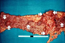
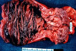
This is a gross photograph of the adenoma from the surgical specimen in this case. Note the large, ulcerated, fungating annular (encircling) carcinoma (1) with areas of hemorrhage (2). Also note the adenomatous polyps (3).
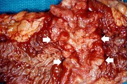
This is a closer view of the previous image demonstrating the raised, annular carcinoma (arrows).
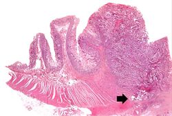
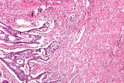
This photomicrograph of the surgical specimen shows the transition between normal mucosa on the left and carcinoma which is invading the wall of the bowel (arrow).
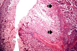
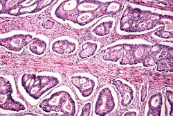
This is a higher-power photomicrograph of the area of transition between the normal (1) and the neoplastic (2) epithelium.
This is a high-power photomicrograph of tumor cells invading the underlying muscularis.
This is a high-power photomicrograph of tumor cells forming glands.
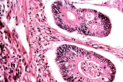
This is a high-power photomicrograph of tumor cells forming glands.
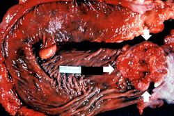
This gross photograph from another case demonstrates an ulcerated adenocarcinoma (arrows) at the rectosigmoid junction.
This is a segment of distal colon from another case. Note the annular tumor that severely compromises the lumen of the colon. There is dilation of the colon proximal to the tumor.


Virtual Microscopy[edit]
Study Questions[edit]
Additional Resources[edit]
Reference[edit]
- eMedicine Medical Library: Imaging in Adenocarcinoma of the Colon
- eMedicine Medical Library: Colon Adenocarcinoma
- Merck Manual: Colorectal Cancer
Journal Articles[edit]
- Yuen ST, Wong MP, Chung LP, Chan SY, Cheung N, Ho J, Leung SY. Up-regulation of lysozyme production in colonic adenomas and adenocarcinomas. Histopathology 1998 Feb;32(2):126-32.
- Strum WB. Colorectal Adenomas. NEJM 2016 March 17 374:1065-1075.
Images[edit]
Related IPLab Cases[edit]
- Lab 7: Lung & Liver: Metastatic Adenocarcinoma
- Lab 7: Lip: Squamous Cell Carcinoma
- Lab 7: Esophagus: Squamous Cell Carcinoma
- Lab 7: Breast: Infiltrating Ductal Carcinoma
- Lab 7: Lung: Bronchogenic Carcinoma
Melena is the passage of digested blood in the feces.