IPLab:Lab 6:Tuberculosis
Contents
Clinical SummaryEdit
During the course of a routine physical examination two months prior to admission, this 57-year-old white male was noted to have a lesion in the upper lobe of the right lung. Initially, he was treated for two weeks with ampicillin. He was then admitted to an outside hospital for further study. All studies including sputum studies for tubercle bacilli, bronchial washings, and bronchoscopy were negative and he was discharged. Review of systems revealed the presence of mild dyspnea on exertion, accompanied by a slightly productive cough. Of interest was the fact that the patient had been PPD positive for the past 4 to 5 years, but this had never been evaluated. On this hospital admission, physical and laboratory examinations were negative. Radiographic examination of the chest revealed a 2 x 2-cm density in the right lower lung field. Several small cavities were identified in this area on CT scan.
The patient underwent a thoracotomy, at which time a portion of the upper lobe of the right lung was removed. Examination of the cut surface revealed small white nodules measuring up to 0.2 cm in diameter.
ImagesEdit
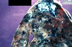
This is a photograph of a section of lung with an apical lesion. This lesion represents an old healed lesion from Mycobacterium tuberculosis infection.
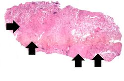
This is a low-power photomicrograph of lung tissue with multiple circumscribed nodules - granulomas (arrows).
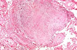
This is a higher-power photomicrograph of a TB granuloma. Note the eosinophilic material in the center of this granuloma (caseous necrosis) and the epithelioid macrophages and giant cells around the periphery.
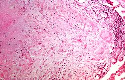
This is a higher-power photomicrograph of a TB granuloma. The area of caseous necrosis is on the left side of the image, there are multinucleated giant cells and epithelioid macrophages throughout the remainder of the tissue.
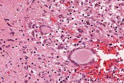
High-power photomicrograph of a TB granuloma with multinucleated giant cells adjacent to an area of caseous necrosis (to the left).
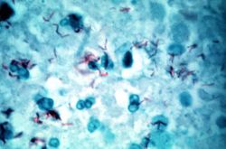
This is a high-power (oil immersion) photomicrograph of granuloma stained with an acid-fast stain. Mycobacterium tuberculosis bacilli stain red.


Virtual MicroscopyEdit
Study QuestionsEdit
Additional ResourcesEdit
ReferenceEdit
Journal ArticlesEdit
- Rodrigues DS, Medeiros EA, Weckx LY, Bonnez W, Salomão R, Kallas EG. Immunophenotypic characterization of peripheral T lymphocytes in Mycobacterium tuberculosis infection and disease. Clin Exp Immunol 2002 Apr;128(1):149-54.
ImagesEdit
Related IPLab CasesEdit
Mycobateria grow very slowly on culture plates, with cultures requiring up to 6 weeks for a positive finding. In lieu of cultures, a more rapid diagnostic test is the PPD--purified protein derivative of tuberculosis--test. PPD is injected under the skin of an individual and then the area is reexamined in 48-72 hours for signs of an inflammatory reaction. A positive test indicates previous exposure to M. tuberculosis.
A thoracotomy is a surgical procedure in which an opening is made in the chest wall.
Caseous means cheesy.
A normal PaCO2 is 35 to 45 mmHg.