IPLab:Lab 6:Rheumatoid Arthritis
Contents
Clinical Summary[edit]
This 57-year-old white female had suffered from rheumatoid arthritis for 20 years. During this period, many joints were involved, some seriously. Because of the severe pain of this arthritis the patient was placed on steroids and was given analgesics, some of which contained acetaminophen. The patient also took additional analgesics (aspirin and/or acetaminophen) to help control the pain. The patient was admitted to the emergency room for weakness and hematemesis. On admission the patient's hematocrit was 21%. Endoscopy demonstrated a large bleeding ulcer and fresh blood in the stomach and proximal duodenum. The sites of bleeding were cauterized; however, shortly after the procedure the patient became hypotensive and died despite aggressive resuscitation.
Autopsy Findings[edit]
There were numerous erosions and ulcers in the gastrointestinal tract and a large quantity of fresh blood in the gastrointestinal tract. There was also significant swelling and deformation in multiple joints. On the medial aspect of the right foot there was a firm, irregular, rubbery subcutaneous nodule measuring 2 x 1.5 cm. The cut surface was whitish-yellow and fibrous.
Images[edit]
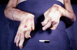
This is a gross photograph of the patient's hands at autopsy. Note the swollen joints and the deforming arthritis.
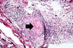
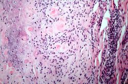
This is a medium-power photomicrograph of the joint capsule surrounding the metacarpal joints. Note the thickening of the capsule and the focal accumulation of inflammatory cells surrounding a central area of fibrinoid necrosis (arrow).
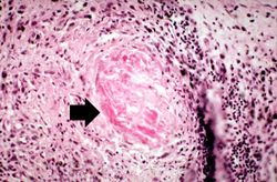
This is a high-power photomicrograph of the joint capsule with another granuloma surrounding a central area of fibrinoid necrosis (arrow).
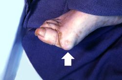
This is a gross photograph of the foot from this same patient. Note the subcutaneous nodule on the medial aspect of the foot (arrow).
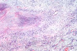
This is a low-power photomicrograph of the subcutaneous nodule from this patient.
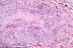
This higher-power photomicrograph of the subcutaneous nodule shows a granulomatous lesion with a necrotic center and a peripheral rim of macrophages, fibrocytes, and occasional lymphocytes. In the necrotic center of the granuloma there is some mineralization (basophilic material).
This higher-power photomicrograph of the subcutaneous nodule again demonstrates the necrotic center and peripheral rim of macrophages, fibrocytes, and occasional lymphocytes. There are focal accumulations of hyaline material (fibrinoid material) within the granuloma.
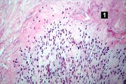
This higher-power photomicrograph of the tissue illustrates the palisading nuclei of the monocytes which are located around the periphery of the central necrotic region (1).
This is a high-power photomicrograph of the mononuclear cells which surround the central area of necrosis. The focal accumulations of fibrinoid material are clearly visible. Lymphocytes are present in the extreme right of this image.
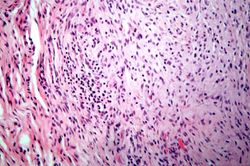
This is a high-power photomicrograph of another region with macrophages (right), fibrocytes (left), and occasional lymphocytes throughout the lesion.


Virtual Microscopy[edit]
Study Questions[edit]
Additional Resources[edit]
Reference[edit]
- eMedicine Medical Library: Rheumatoid Arthritis
- Merck Manual: Rheumatoid Arthritis (RA)
- National Library of Medicine: Rheumatoid Arthritis
Journal Articles[edit]
- Carter RA, O'Donnell K, Sachthep S, Cicuttini F, Boyd AW, Wicks IP. Characterization of a human synovial cell antigen: VCAM-1 and inflammatory arthritis. Immunol Cell Biol 2001 Oct;79(5):419-28.
Images[edit]
Related IPLab Cases[edit]
Hematemesis is the vomiting of blood.
A hematocrit value represents the number of packed red cells in mL per 100 mL of centrifuged whole blood--expressed as a percentage. A normal hematocrit for a female is 34 to 44%.
Autoimmune disorders involve an immune response directed at the host's own cells.