Difference between revisions of "IPLab:Lab 11:Cysticercosis"
Seung Park (talk | contribs) (→Images) |
(→Clinical Summary) |
||
| (One intermediate revision by the same user not shown) | |||
| Line 2: | Line 2: | ||
This 29-year-old woman was admitted to the hospital because of repeated tonic-clonic seizures. The patient was a tour guide leading groups of tourist to Tibet for two-month walking/camping tours in the Himalayas. Her seizures were easily controlled by intravenous administration of phenytoin. The WBC count was 13,000, with 5% eosinophils and the erythrocyte sedimentation rate was slightly elevated. A cranial CT performed with and without contrast revealed two ring-enhancing lesions. The patient underwent a craniotomy and excisional biopsy. | This 29-year-old woman was admitted to the hospital because of repeated tonic-clonic seizures. The patient was a tour guide leading groups of tourist to Tibet for two-month walking/camping tours in the Himalayas. Her seizures were easily controlled by intravenous administration of phenytoin. The WBC count was 13,000, with 5% eosinophils and the erythrocyte sedimentation rate was slightly elevated. A cranial CT performed with and without contrast revealed two ring-enhancing lesions. The patient underwent a craniotomy and excisional biopsy. | ||
| − | + | Histopathologic exam of the surgical specimen revealed a capsule of dense connective tissue surrounding a cavity that contained a partially degenerated scolex of Taenia solium. | |
| − | |||
| − | Histopathologic exam revealed a capsule of dense connective tissue surrounding a cavity that contained a partially degenerated scolex of Taenia solium. | ||
== Images == | == Images == | ||
Latest revision as of 22:03, 9 July 2020
Contents
Clinical SummaryEdit
This 29-year-old woman was admitted to the hospital because of repeated tonic-clonic seizures. The patient was a tour guide leading groups of tourist to Tibet for two-month walking/camping tours in the Himalayas. Her seizures were easily controlled by intravenous administration of phenytoin. The WBC count was 13,000, with 5% eosinophils and the erythrocyte sedimentation rate was slightly elevated. A cranial CT performed with and without contrast revealed two ring-enhancing lesions. The patient underwent a craniotomy and excisional biopsy.
Histopathologic exam of the surgical specimen revealed a capsule of dense connective tissue surrounding a cavity that contained a partially degenerated scolex of Taenia solium.
ImagesEdit
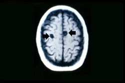
This is a head CT showing the two cysts (arrows).
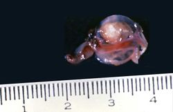
This is a photograph of the cyst that was surgically removed. The cyst is filled with a clear fluid and contains a scolex.

This photograph of an autopsy specimen from another patient shows an adult tapeworm in the intestine. Note that the worm attaches to the luminal surface of the intestine via the scolex.
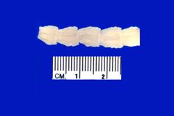
This is a photograph of the body of an adult tapeworm. Note the body segments.


Study QuestionsEdit
Additional ResourcesEdit
ReferenceEdit
- eMedicine Medical Library: Cysticercosis
- eMedicine Medical Library: Cysticercosis in Emergency Medicine
- Merck Manual: Taeniasis Solium and Cysticercosis
Journal ArticlesEdit
- Garcia HH, Del Brutto OH. Taenia solium cysticercosis. Infect Dis Clin North Am 2000 Mar;14(1):97-119, ix.
ImagesEdit
A tonic-clonic seizure involves loss of consciousness followed by tonic, then clonic, convulsions.
A normal white blood cell count is 4000-11,000 cells/mm³.
An elevated erythrocyte sedimentation rate is a non-specific indicator of inflammation.