File:IPLab6Scleroderma5.jpg
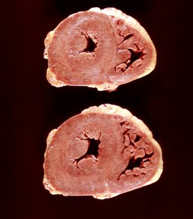
Revision as of 19:59, 20 August 2013 by Peter Anderson (talk | contribs) (This is a gross photograph of the heart from this case. There is thickening of the left ventricular wall and some thickening of the right ventricle as well.)
No higher resolution available.
IPLab6Scleroderma5.jpg (398 × 450 pixels, file size: 19 KB, MIME type: image/jpeg)
This is a gross photograph of the heart from this case. There is thickening of the left ventricular wall and some thickening of the right ventricle as well.
File history
Click on a date/time to view the file as it appeared at that time.
| Date/Time | Thumbnail | Dimensions | User | Comment | |
|---|---|---|---|---|---|
| current | 19:59, 20 August 2013 | | 398 × 450 (19 KB) | Peter Anderson (talk | contribs) | This is a gross photograph of the heart from this case. There is thickening of the left ventricular wall and some thickening of the right ventricle as well. |
- You cannot overwrite this file.
File usage
The following page links to this file: