File:IPLab5Gout5.jpg
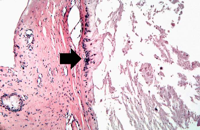
Revision as of 15:19, 20 August 2013 by Peter Anderson (talk | contribs) (This is a higher-power photomicrograph of the edge of the tophus. Most of the urate crystals dissolve away during processing. The inflammatory cells at the edge of these foci are clearly visible (arrow).)
No higher resolution available.
IPLab5Gout5.jpg (688 × 450 pixels, file size: 60 KB, MIME type: image/jpeg)
This is a higher-power photomicrograph of the edge of the tophus. Most of the urate crystals dissolve away during processing. The inflammatory cells at the edge of these foci are clearly visible (arrow).
A tophus is a chalky accumulation of urate crystals found in the tissue surrounding a joint.
File history
Click on a date/time to view the file as it appeared at that time.
| Date/Time | Thumbnail | Dimensions | User | Comment | |
|---|---|---|---|---|---|
| current | 15:19, 20 August 2013 | | 688 × 450 (60 KB) | Peter Anderson (talk | contribs) | This is a higher-power photomicrograph of the edge of the tophus. Most of the urate crystals dissolve away during processing. The inflammatory cells at the edge of these foci are clearly visible (arrow). |
- You cannot overwrite this file.
File usage
The following page links to this file: