File:IPLab5Gout1.jpg
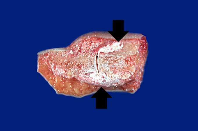
Revision as of 15:17, 20 August 2013 by Peter Anderson (talk | contribs) (This is a gross photograph of an index finger from a patient with gout. The finger has been sectioned longitudinally to demonstrate the distal interphalangeal joint. Note the white chalky material within and adjacent to the joint (arrows).)
No higher resolution available.
IPLab5Gout1.jpg (681 × 450 pixels, file size: 22 KB, MIME type: image/jpeg)
This is a gross photograph of an index finger from a patient with gout. The finger has been sectioned longitudinally to demonstrate the distal interphalangeal joint. Note the white chalky material within and adjacent to the joint (arrows).
File history
Click on a date/time to view the file as it appeared at that time.
| Date/Time | Thumbnail | Dimensions | User | Comment | |
|---|---|---|---|---|---|
| current | 15:17, 20 August 2013 | | 681 × 450 (22 KB) | Peter Anderson (talk | contribs) | This is a gross photograph of an index finger from a patient with gout. The finger has been sectioned longitudinally to demonstrate the distal interphalangeal joint. Note the white chalky material within and adjacent to the joint (arrows). |
- You cannot overwrite this file.
File usage
The following page links to this file: