File:IPLab5Antitrypsin2.jpg
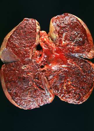
Revision as of 18:07, 19 August 2013 by Peter Anderson (talk | contribs) (This is a gross photograph of the cut sections of lung from this case. The lung parenchyma is markedly hemorrhagic and consolidated. Again the hemorrhage makes it difficult to appreciate the emphysematous changes.)
No higher resolution available.
IPLab5Antitrypsin2.jpg (323 × 450 pixels, file size: 32 KB, MIME type: image/jpeg)
This is a gross photograph of the cut sections of lung from this case. The lung parenchyma is markedly hemorrhagic and consolidated. Again the hemorrhage makes it difficult to appreciate the emphysematous changes.
Consolidation is the filling of lung air spaces with exudate--this is a sign of pneumonia.
File history
Click on a date/time to view the file as it appeared at that time.
| Date/Time | Thumbnail | Dimensions | User | Comment | |
|---|---|---|---|---|---|
| current | 18:07, 19 August 2013 | | 323 × 450 (32 KB) | Peter Anderson (talk | contribs) | This is a gross photograph of the cut sections of lung from this case. The lung parenchyma is markedly hemorrhagic and consolidated. Again the hemorrhage makes it difficult to appreciate the emphysematous changes. |
- You cannot overwrite this file.
File usage
There are no pages that link to this file.