File:IPLab4AtheromatousEmboli3.jpg
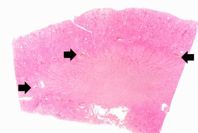
Revision as of 17:00, 19 August 2013 by Seung Park (talk | contribs) (This is a low-power photomicrograph of kidney tissue. Several blood vessels can be identified at the corticomedullary junction (arrows).)
No higher resolution available.
IPLab4AtheromatousEmboli3.jpg (671 × 450 pixels, file size: 34 KB, MIME type: image/jpeg)
This is a low-power photomicrograph of kidney tissue. Several blood vessels can be identified at the corticomedullary junction (arrows).
File history
Click on a date/time to view the file as it appeared at that time.
| Date/Time | Thumbnail | Dimensions | User | Comment | |
|---|---|---|---|---|---|
| current | 17:00, 19 August 2013 | | 671 × 450 (34 KB) | Seung Park (talk | contribs) | This is a low-power photomicrograph of kidney tissue. Several blood vessels can be identified at the corticomedullary junction (arrows). |
- You cannot overwrite this file.
File usage
The following page links to this file: