File:IPLab3Tuberculosis2.jpg
Revision as of 03:38, 19 August 2013 by Seung Park (talk | contribs) (This low-power photomicrograph of a section of lung reveals multiple large nodules (1) with pale eosinophilic centers surrounded by a rim of blue-staining nuclei. In addition to the large nodules, there are several smaller nodules throughout the slide ...)
No higher resolution available.
IPLab3Tuberculosis2.jpg (675 × 450 pixels, file size: 22 KB, MIME type: image/jpeg)
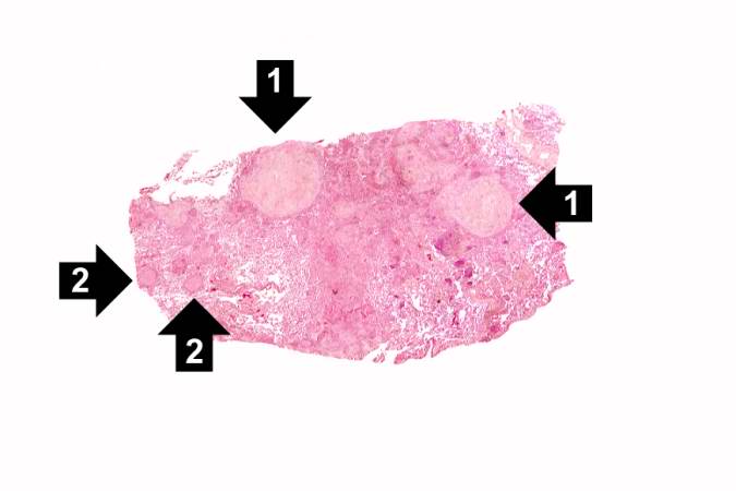
This low-power photomicrograph of a section of lung reveals multiple large nodules (1) with pale eosinophilic centers surrounded by a rim of blue-staining nuclei. In addition to the large nodules, there are several smaller nodules throughout the slide (2).
File history
Click on a date/time to view the file as it appeared at that time.
| Date/Time | Thumbnail | Dimensions | User | Comment | |
|---|---|---|---|---|---|
| current | 03:38, 19 August 2013 | | 675 × 450 (22 KB) | Seung Park (talk | contribs) | This low-power photomicrograph of a section of lung reveals multiple large nodules (1) with pale eosinophilic centers surrounded by a rim of blue-staining nuclei. In addition to the large nodules, there are several smaller nodules throughout the slide ... |
- You cannot overwrite this file.
File usage
The following page links to this file: