File:IPLab3LobarPneumonia2.jpg
Revision as of 03:16, 19 August 2013 by Seung Park (talk | contribs) (This is a cut section of a lung from the preceding image. Note the whitish discoloration of the lung tissue in the upper lobe (arrows) compared to the normal collapsed and pink staining lung lobe in the left-hand portion of the photograph. The white di...)
No higher resolution available.
IPLab3LobarPneumonia2.jpg (679 × 450 pixels, file size: 55 KB, MIME type: image/jpeg)
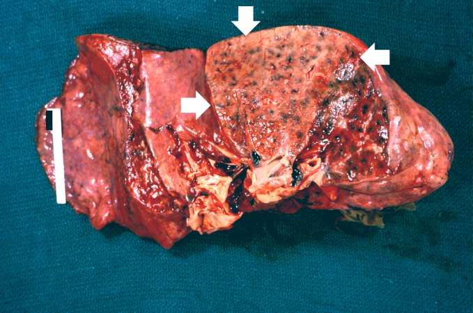
This is a cut section of a lung from the preceding image. Note the whitish discoloration of the lung tissue in the upper lobe (arrows) compared to the normal collapsed and pink staining lung lobe in the left-hand portion of the photograph. The white discoloration in this tissue is due to infiltration of leukocytes (primarily neutrophils). Note that only one lobe of the lung is involved in this patient with lobar pneumonia.
In alcoholics, aspiration pneumonia is common--bacteria enter the lung via aspiration of gastric contents.
File history
Click on a date/time to view the file as it appeared at that time.
| Date/Time | Thumbnail | Dimensions | User | Comment | |
|---|---|---|---|---|---|
| current | 03:16, 19 August 2013 | | 679 × 450 (55 KB) | Seung Park (talk | contribs) | This is a cut section of a lung from the preceding image. Note the whitish discoloration of the lung tissue in the upper lobe (arrows) compared to the normal collapsed and pink staining lung lobe in the left-hand portion of the photograph. The white di... |
- You cannot overwrite this file.
File usage
The following page links to this file: