File:IPLab3HealedMyocardialInfarction4.jpg
Revision as of 04:37, 19 August 2013 by Seung Park (talk | contribs) (This is another high-power photomicrograph of a healed myocardial infarction. Note the remaining normal myocytes (1), the fibrous connective tissue (2), and occasional hypereosinophilic myocytes indicating recent acute ischemic injury (arrow).)
No higher resolution available.
IPLab3HealedMyocardialInfarction4.jpg (693 × 450 pixels, file size: 61 KB, MIME type: image/jpeg)
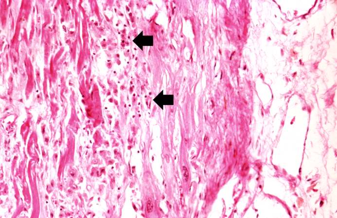
This is another high-power photomicrograph of a healed myocardial infarction. Note the remaining normal myocytes (1), the fibrous connective tissue (2), and occasional hypereosinophilic myocytes indicating recent acute ischemic injury (arrow).
Myocardial infarction is necrosis of myocardial tissue which occurs as a result of a deprivation of blood supply, and thus oxygen, to the heart tissue. Blockage of blood supply to the myocardium is caused by occlusion of a coronary artery.
File history
Click on a date/time to view the file as it appeared at that time.
| Date/Time | Thumbnail | Dimensions | User | Comment | |
|---|---|---|---|---|---|
| current | 04:37, 19 August 2013 | | 693 × 450 (61 KB) | Seung Park (talk | contribs) | This is another high-power photomicrograph of a healed myocardial infarction. Note the remaining normal myocytes (1), the fibrous connective tissue (2), and occasional hypereosinophilic myocytes indicating recent acute ischemic injury (arrow). |
- You cannot overwrite this file.
File usage
There are no pages that link to this file.