File:IPLab3ChronicPepticUlcer9.jpg
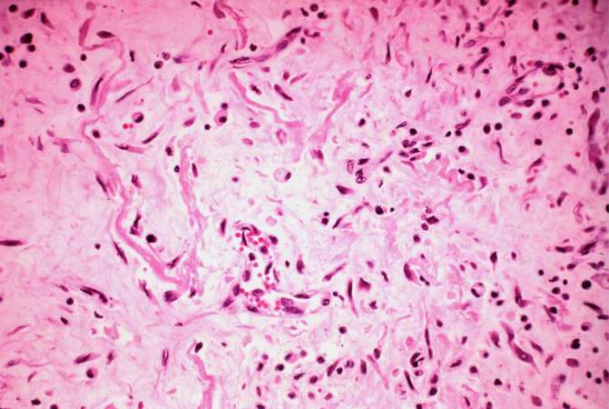
Revision as of 04:17, 19 August 2013 by Seung Park (talk | contribs) (This high-power photomicrograph demonstrates the granulation tissue within the base of the ulcer.)
No higher resolution available.
IPLab3ChronicPepticUlcer9.jpg (671 × 450 pixels, file size: 50 KB, MIME type: image/jpeg)
This high-power photomicrograph demonstrates the granulation tissue within the base of the ulcer.
File history
Click on a date/time to view the file as it appeared at that time.
| Date/Time | Thumbnail | Dimensions | User | Comment | |
|---|---|---|---|---|---|
| current | 04:17, 19 August 2013 | | 671 × 450 (50 KB) | Seung Park (talk | contribs) | This high-power photomicrograph demonstrates the granulation tissue within the base of the ulcer. |
- You cannot overwrite this file.
File usage
There are no pages that link to this file.