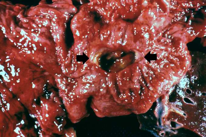
File:IPLab3ChronicPepticUlcer1.jpg
Revision as of 04:14, 19 August 2013 by Seung Park (talk | contribs) (This is a gross photograph of a stomach containing an ulcer. Note the folded pink gastric mucosa that extends up to the edge of the ulcer (arrows).)
No higher resolution available.
IPLab3ChronicPepticUlcer1.jpg (679 × 450 pixels, file size: 58 KB, MIME type: image/jpeg)
This is a gross photograph of a stomach containing an ulcer. Note the folded pink gastric mucosa that extends up to the edge of the ulcer (arrows).
File history
Click on a date/time to view the file as it appeared at that time.
| Date/Time | Thumbnail | Dimensions | User | Comment | |
|---|---|---|---|---|---|
| current | 04:14, 19 August 2013 | | 679 × 450 (58 KB) | Seung Park (talk | contribs) | This is a gross photograph of a stomach containing an ulcer. Note the folded pink gastric mucosa that extends up to the edge of the ulcer (arrows). |
- You cannot overwrite this file.
File usage
There are no pages that link to this file.