File:IPLab2Metaplasia6.jpg
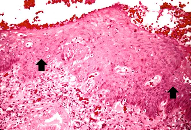
Revision as of 15:45, 19 August 2013 by Peter Anderson (talk | contribs) (A high-power photomicrograph of the squamous epithelium shows inflammatory cells in the subepithelial tissue and the formation of keratinized epithelium (arrows).)
No higher resolution available.
IPLab2Metaplasia6.jpg (663 × 450 pixels, file size: 67 KB, MIME type: image/jpeg)
A high-power photomicrograph of the squamous epithelium shows inflammatory cells in the subepithelial tissue and the formation of keratinized epithelium (arrows).
File history
Click on a date/time to view the file as it appeared at that time.
| Date/Time | Thumbnail | Dimensions | User | Comment | |
|---|---|---|---|---|---|
| current | 15:45, 19 August 2013 | | 663 × 450 (67 KB) | Peter Anderson (talk | contribs) | A high-power photomicrograph of the squamous epithelium shows inflammatory cells in the subepithelial tissue and the formation of keratinized epithelium (arrows). |
- You cannot overwrite this file.
File usage
There are no pages that link to this file.