File:IPLab1Tuberculosis8.jpg
Revision as of 18:42, 31 October 2016 by Peter Anderson (talk | contribs) (Peter Anderson uploaded a new version of "File:IPLab1Tuberculosis8.jpg")
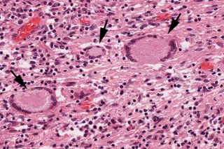
Size of this preview: 800 × 533 pixels. Other resolutions: 320 × 213 pixels | 1,386 × 924 pixels.
Original file (1,386 × 924 pixels, file size: 246 KB, MIME type: image/jpeg)
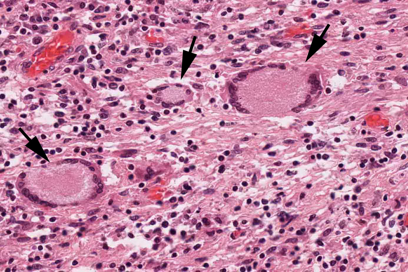
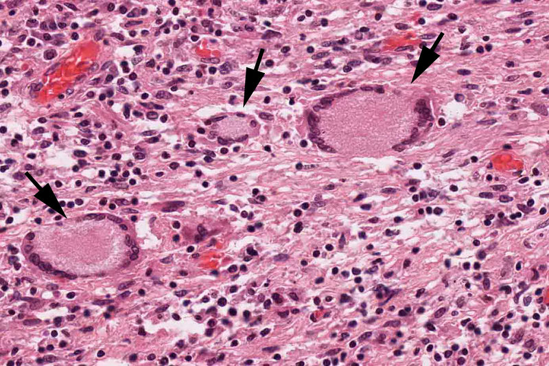
This is a high-power photomicrograph of the Langhans-type multinucleated giant cell which is characteristic of tuberculous granulomas (arrow). Note the horseshoe shape of the nuclei in this giant cell. The majority of the cells in the upper left portion of this section are macrophages which provide the major cellular component in a granuloma. Note the smaller number of small blue-staining cells in the peripheral portions of this granuloma to the left of which are lymphocytes.
File history
Click on a date/time to view the file as it appeared at that time.
| Date/Time | Thumbnail | Dimensions | User | Comment | |
|---|---|---|---|---|---|
| current | 18:42, 31 October 2016 | | 1,386 × 924 (246 KB) | Peter Anderson (talk | contribs) |
- You cannot overwrite this file.
File usage
The following file is a duplicate of this file (more details):
There are no pages that link to this file.