File:IPLab13Meningococcemia3.jpg
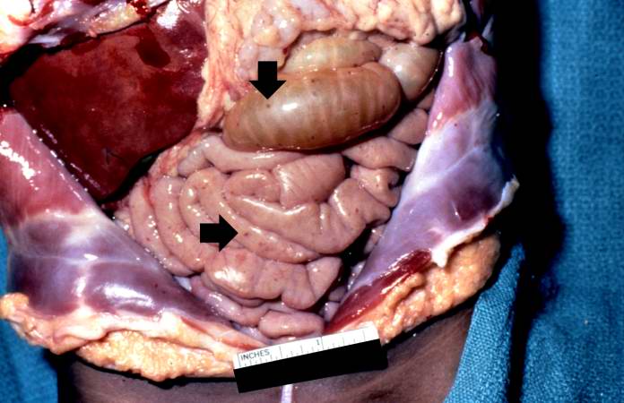
Revision as of 06:05, 21 August 2013 by Seung Park (talk | contribs) (In this gross photograph of the abdomen taken at the autopsy, there are petechial hemorrhages on the viscera (arrows).)
No higher resolution available.
IPLab13Meningococcemia3.jpg (696 × 450 pixels, file size: 48 KB, MIME type: image/jpeg)
In this gross photograph of the abdomen taken at the autopsy, there are petechial hemorrhages on the viscera (arrows).
File history
Click on a date/time to view the file as it appeared at that time.
| Date/Time | Thumbnail | Dimensions | User | Comment | |
|---|---|---|---|---|---|
| current | 06:05, 21 August 2013 | | 696 × 450 (48 KB) | Seung Park (talk | contribs) | In this gross photograph of the abdomen taken at the autopsy, there are petechial hemorrhages on the viscera (arrows). |
- You cannot overwrite this file.
File usage
The following page links to this file: