File:IPLab13Hyaline8.jpg
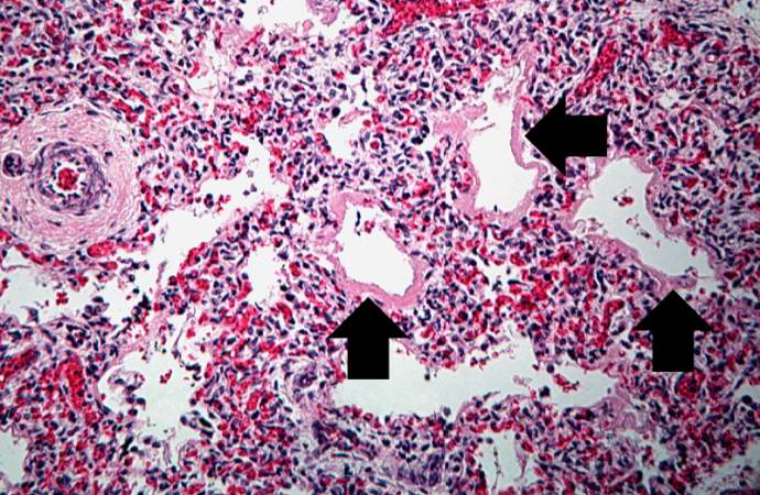
Revision as of 05:52, 21 August 2013 by Seung Park (talk | contribs) (This medium-power photomicrograph shows the pink acellular homogeneous material lining the alveoli which comprises the hyaline membranes (arrows). The interstitium shows congestion, as in previous sections.)
No higher resolution available.
IPLab13Hyaline8.jpg (690 × 450 pixels, file size: 90 KB, MIME type: image/jpeg)
This medium-power photomicrograph shows the pink acellular homogeneous material lining the alveoli which comprises the hyaline membranes (arrows). The interstitium shows congestion, as in previous sections.
File history
Click on a date/time to view the file as it appeared at that time.
| Date/Time | Thumbnail | Dimensions | User | Comment | |
|---|---|---|---|---|---|
| current | 05:52, 21 August 2013 | | 690 × 450 (90 KB) | Seung Park (talk | contribs) | This medium-power photomicrograph shows the pink acellular homogeneous material lining the alveoli which comprises the hyaline membranes (arrows). The interstitium shows congestion, as in previous sections. |
- You cannot overwrite this file.
File usage
The following page links to this file: