File:IPLab13Hyaline6.jpg
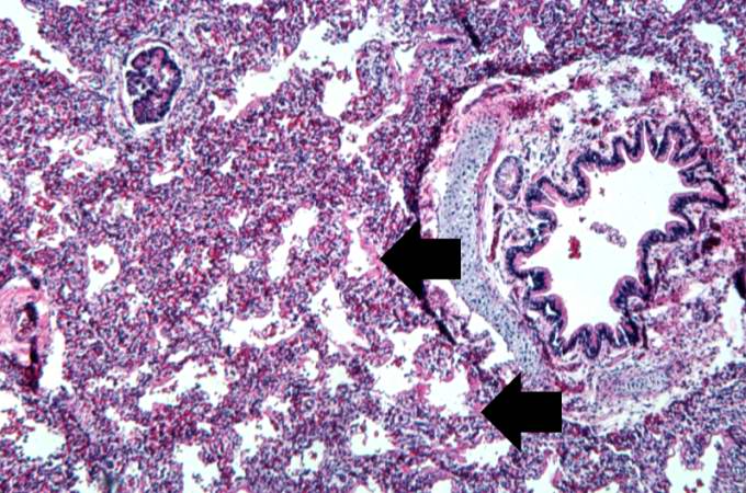
Revision as of 05:51, 21 August 2013 by Seung Park (talk | contribs) (This is a medium-power photomicrograph showing a large bronchus with cartilage. Interstitial congestion with numerous red cells is apparent. Even at this magnification hyaline membranes (arrows) can be seen lining the alveoli.)
No higher resolution available.
IPLab13Hyaline6.jpg (680 × 450 pixels, file size: 85 KB, MIME type: image/jpeg)
This is a medium-power photomicrograph showing a large bronchus with cartilage. Interstitial congestion with numerous red cells is apparent. Even at this magnification hyaline membranes (arrows) can be seen lining the alveoli.
File history
Click on a date/time to view the file as it appeared at that time.
| Date/Time | Thumbnail | Dimensions | User | Comment | |
|---|---|---|---|---|---|
| current | 05:51, 21 August 2013 | | 680 × 450 (85 KB) | Seung Park (talk | contribs) | This is a medium-power photomicrograph showing a large bronchus with cartilage. Interstitial congestion with numerous red cells is apparent. Even at this magnification hyaline membranes (arrows) can be seen lining the alveoli. |
- You cannot overwrite this file.
File usage
The following page links to this file: