Difference between revisions of "Cytologically Yours: CoW: 20140220"
(→Cytology) |
|||
| Line 22: | Line 22: | ||
CytologicallyYoursCoW20140220Cytology1.jpg|20x magnification of a cellular specimen with numerous follicles and absent colloid. | CytologicallyYoursCoW20140220Cytology1.jpg|20x magnification of a cellular specimen with numerous follicles and absent colloid. | ||
CytologicallyYoursCoW20140220Cytology2.jpg|40x magnification with follicular cells forming microfollicular groups and inspissated colloid. | CytologicallyYoursCoW20140220Cytology2.jpg|40x magnification with follicular cells forming microfollicular groups and inspissated colloid. | ||
| − | CytologicallyYoursCoW20140220Cytology3.jpg|60x magnification with follicular cells forming microfollicular groups and inspissated colloid. | + | CytologicallyYoursCoW20140220Cytology3.jpg|60x magnification with follicular cells forming microfollicular groups and inspissated colloid. Nuclear are crowded with overlapping. The nuclei are enlarged. |
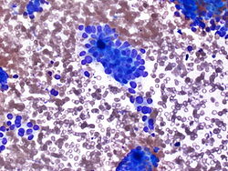
| − | CytologicallyYoursCoW20140220Cytology4.jpg|40x magnification with follicular cells forming microfollicular | + | CytologicallyYoursCoW20140220Cytology4.jpg|40x magnification with follicular cells forming a microfollicular group and inspissated colloid. |
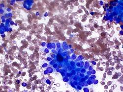
CytologicallyYoursCoW20140220Cytology5.jpg|40x magnification of Pap stained material showing magnification with follicular cells forming microfollicular groups and inspissated colloid.. | CytologicallyYoursCoW20140220Cytology5.jpg|40x magnification of Pap stained material showing magnification with follicular cells forming microfollicular groups and inspissated colloid.. | ||
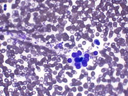
CytologicallyYoursCoW20140220Cytology6.jpg|60x magnification of Pap stained material with microfollicles. | CytologicallyYoursCoW20140220Cytology6.jpg|60x magnification of Pap stained material with microfollicles. | ||
Latest revision as of 22:47, 4 March 2014
Contents
Clinical Summary
The patient is a 58 year old male with back and neck pain.
Past Medical History
- Cervical spondylosis
- Hypertension
- Hyperlipidemia
Past Surgical History
- No surgical history
Radiology
- Ultrasound of the neck shows a solid interpolar nodule 1.8 x 1.6 cm in the left lobe of the thyroid.
Clinical Plan
Fine needle aspiration of the nodule is scheduled.
Pathology
Cytology
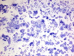
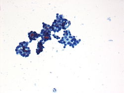
20x magnification of a cellular specimen with numerous follicles and absent colloid.
40x magnification with follicular cells forming microfollicular groups and inspissated colloid.
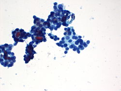
60x magnification with follicular cells forming microfollicular groups and inspissated colloid. Nuclear are crowded with overlapping. The nuclei are enlarged.
40x magnification with follicular cells forming a microfollicular group and inspissated colloid.
40x magnification of Pap stained material showing magnification with follicular cells forming microfollicular groups and inspissated colloid..
60x magnification of Pap stained material with microfollicles.


Resident Questions
Final Diagnosis
Cytology
- Follicular neoplasm.
Discussion
The diagnostic criteria for follicular lesions are based on cellularity and the presence of colloid. Follicular lesions will be cellular with an absence of the variability in the size of the follicular groups, with a predominately microfollicular pattern. Colloid will be scant to absent, except for the presence of inspissated colloid in the center of follicular groups. There is often a disorganized, crowded follicular pattern with some nucleomegaly and nucleoli.
| ||||||||