Difference between revisions of "Cytologically Yours: CoW: 20140213"
| Line 31: | Line 31: | ||
====Resident Questions==== | ====Resident Questions==== | ||
| − | * <spoiler text="What | + | * <spoiler text="What was the initial diagnosis on rapid interpretation?"> Adenocarcinoma. </spoiler> |
| − | |||
| − | |||
| − | |||
| − | |||
| − | |||
<div class="usermessage mw-customtoggle-diagnosis" style="cursor:pointer">Click here to toggle the diagnosis and discussion.</div> | <div class="usermessage mw-customtoggle-diagnosis" style="cursor:pointer">Click here to toggle the diagnosis and discussion.</div> | ||
| Line 48: | Line 43: | ||
| − | + | ===Cell Block=== | |
| + | <gallery heights="250px" widths="250px"> | ||
| + | CytologicallyYoursCoW20140213Biopsy1.jpg|40x magnification of cell block of pancreatic mass showing small glands with macronucleoli. | ||
</gallery> | </gallery> | ||
Revision as of 22:14, 4 March 2014
Contents
Clinical Summary
The patient is a 66yo old male presenting with jaundice.
Past Medical History
- History of prostate cancer 5 years ago.
- Hyperlipidemia
- Chronic back pain
Past Surgical History
- Prostatectomy (2009)
- Thyroidectomy (2004)
Ultrasound
- Ultrasound of head of pancreas shows a 43mm x 23mm ill defined mass.
Clinical Plan
The differential diagnosis included pancreatic adenocarcinoma and metastatic prostate cancer.
Pathology
Cytology
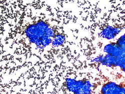
10x magnification of large cohesive groups of cells.
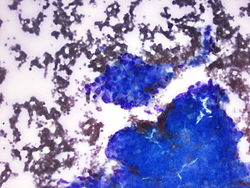
40x magnification showing atypical cells with large irregular nuclei.
20x magnification showing atypical cells with large irregular nuclei.
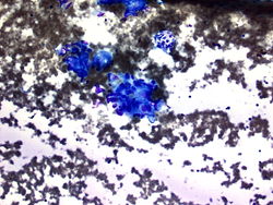
40x magnification showing large, markedly pleomorphic cells.
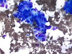
40x magnification of malignant cell groups.
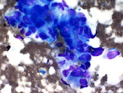
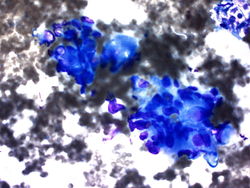
60x magnification of malignant cells. Macronucleoli are easily identified.


Resident Questions
Final Diagnosis
Cytology
- Metastatic prostate carcinoma.
Cell Block
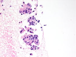
40x magnification of cell block of pancreatic mass showing small glands with macronucleoli.

Biopsy Diagnosis
- Conventional high grade sarcoma, sclerotic type.
Discussion
The experience of FNA of osteosarcoma is mainly with conventional high-grade intramedullary sarcoma and to the rare high-grade surface osteosarcoma. Smears usually contain dissociated neoplastic cells and cell clusters.
| ||||||||