Difference between revisions of "Cytologically Yours: CoW: 20140206"
(Created page with "== Clinical Summary == The patient is a 12 year old female with a six month history of left shoulder pain. The patient had tried Aleve and had several chiropractic visits whi...") |
|||
| Line 19: | Line 19: | ||
<gallery heights="250px" widths="250px"> | <gallery heights="250px" widths="250px"> | ||
CytologicallyYoursCoW20140206Cytology1.jpg|40x magnification of highly atypical malignant appearing cells. | CytologicallyYoursCoW20140206Cytology1.jpg|40x magnification of highly atypical malignant appearing cells. | ||
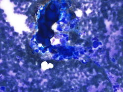
| − | + | CytologicallyYoursCoW20140206Cytology2.jpg|20x magnification showing osteoid formation. | |
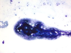
CytologicallyYoursCoW20140206Cytology3.jpg|40x magnification showing osteoid formation and malignant appearing cells. | CytologicallyYoursCoW20140206Cytology3.jpg|40x magnification showing osteoid formation and malignant appearing cells. | ||
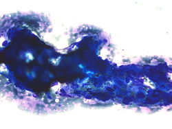
CytologicallyYoursCoW20140206Cytology5.jpg|40x magnification of osteoid. | CytologicallyYoursCoW20140206Cytology5.jpg|40x magnification of osteoid. | ||
Revision as of 21:25, 4 March 2014
Contents
Clinical Summary
The patient is a 12 year old female with a six month history of left shoulder pain. The patient had tried Aleve and had several chiropractic visits which were unsuccessful at relieving the pain.
Past Medical History
- Previously heathy
Past Surgical History
- No surgical history
Radiology
- AP/Lateral images show a destructive and aggressive appearing lesion in the left proximal huerus in the metaphysis extending 7.5cm distally in the diaphysis.
Clinical Plan
The differential diagnosis included osteosarcoma and Ewing sarcoma. MRI and CT guided biopsy are scheduled.
Pathology
Cytology
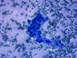
40x magnification of highly atypical malignant appearing cells.
20x magnification showing osteoid formation.
40x magnification showing osteoid formation and malignant appearing cells.
40x magnification of osteoid.


Resident Questions
Final Diagnosis
Cytology
- Positive for malignancy, the differential diagnosis includes melanoma and Hodgkin lymphoma.
Biopsy
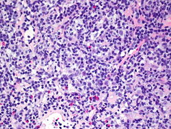
20x magnification of lymph node core biopsy.
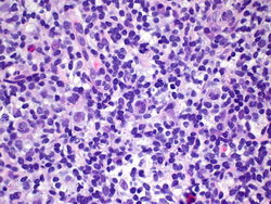
40x magnification of lymph node core biopsy.
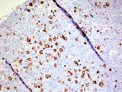
CD 15 with membranous staining.
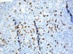
CD 30 with membranous staining.


Biopsy Diagnosis
- Classical Hodgkin lymphoma, favor mixed type.
- CD15 Positive in tumor cells
- CD30 Positive in tumor cells
- PAX5 Weakly positive
- CD20 Positive in background lymphocytes
- CD3 Positive in background lymphocytes
- S100 Negative
- Mart1 Negative
- HMB45 Negative
Discussion
The features of Hodgkin lymphoma include atypical (Hodgkin cells) and Reed-Sternberg cells. The nucleus should be 3-4x the size of a small lymphocyte. In classic Hodgkin lymphoma, scattered eosinophils, plasma cells, histiocytes, and a predominately CD3+ lymphocyte population will be seen in the background. The immunophenotype of classic Hodgkin lymphoma shows CD15, CD30, MUM1, and weak PAX5 positivity. Histology is usually needed to subtype Hodgkin lymphoma.
| ||||||||