File:IPLab9Diphtheria1.jpg
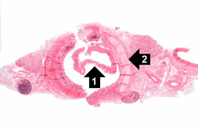
Revision as of 03:49, 21 August 2013 by Seung Park (talk | contribs) (This is a low-power photomicrograph of the trachea with the diphtheritic membrane (1), which has pulled away from the tracheal lining during histological processing. Note the tracheal cartilage (2) present in this section.)
No higher resolution available.
IPLab9Diphtheria1.jpg (685 × 450 pixels, file size: 28 KB, MIME type: image/jpeg)
This is a low-power photomicrograph of the trachea with the diphtheritic membrane (1), which has pulled away from the tracheal lining during histological processing. Note the tracheal cartilage (2) present in this section.
File history
Click on a date/time to view the file as it appeared at that time.
| Date/Time | Thumbnail | Dimensions | User | Comment | |
|---|---|---|---|---|---|
| current | 03:49, 21 August 2013 | | 685 × 450 (28 KB) | Seung Park (talk | contribs) | This is a low-power photomicrograph of the trachea with the diphtheritic membrane (1), which has pulled away from the tracheal lining during histological processing. Note the tracheal cartilage (2) present in this section. |
- You cannot overwrite this file.
File usage
The following page links to this file: