File:IPLab8Polio4.jpg
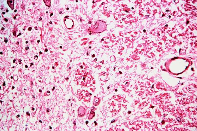
Revision as of 02:44, 21 August 2013 by Seung Park (talk | contribs) (This is a higher-power photomicrograph taken at the junction of the white and gray matter. Note the inflammatory cellular infiltrate and tissue breakdown. There is significant loss of neurons and myelin in this area.)
No higher resolution available.
IPLab8Polio4.jpg (677 × 450 pixels, file size: 77 KB, MIME type: image/jpeg)
This is a higher-power photomicrograph taken at the junction of the white and gray matter. Note the inflammatory cellular infiltrate and tissue breakdown. There is significant loss of neurons and myelin in this area.
An infiltrate is an accumulation of cells in the lung parenchyma--this is a sign of pneumonia.
File history
Click on a date/time to view the file as it appeared at that time.
| Date/Time | Thumbnail | Dimensions | User | Comment | |
|---|---|---|---|---|---|
| current | 02:44, 21 August 2013 | | 677 × 450 (77 KB) | Seung Park (talk | contribs) | This is a higher-power photomicrograph taken at the junction of the white and gray matter. Note the inflammatory cellular infiltrate and tissue breakdown. There is significant loss of neurons and myelin in this area. |
- You cannot overwrite this file.
File usage
The following page links to this file: