File:IPLab6ChronicRejection1.jpg
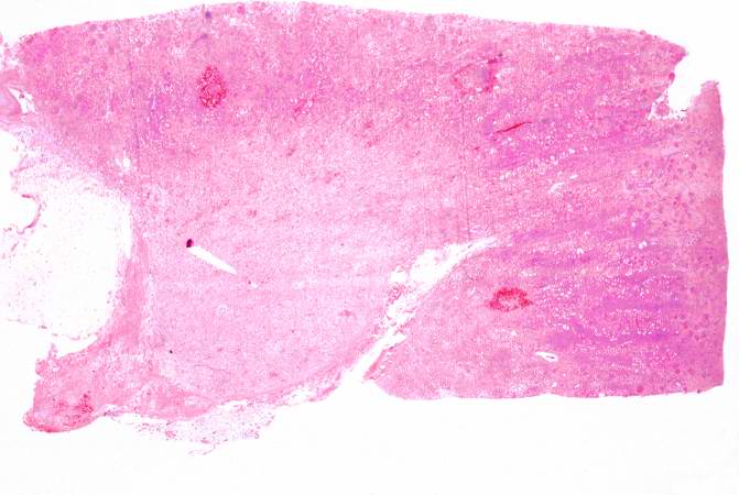
Revision as of 21:48, 20 August 2013 by Seung Park (talk | contribs) (This is a low-power photomicrograph of the kidney from this case of chronic transplant rejection. Note the focal areas of hemorrhage and inflammatory cell infiltrate in this section.)
No higher resolution available.
IPLab6ChronicRejection1.jpg (671 × 450 pixels, file size: 43 KB, MIME type: image/jpeg)
This is a low-power photomicrograph of the kidney from this case of chronic transplant rejection. Note the focal areas of hemorrhage and inflammatory cell infiltrate in this section.
An infiltrate is an accumulation of cells in the lung parenchyma--this is a sign of pneumonia.
File history
Click on a date/time to view the file as it appeared at that time.
| Date/Time | Thumbnail | Dimensions | User | Comment | |
|---|---|---|---|---|---|
| current | 21:48, 20 August 2013 | | 671 × 450 (43 KB) | Seung Park (talk | contribs) | This is a low-power photomicrograph of the kidney from this case of chronic transplant rejection. Note the focal areas of hemorrhage and inflammatory cell infiltrate in this section. |
- You cannot overwrite this file.
File usage
There are no pages that link to this file.