File:IPLab6Hashimoto4.jpg
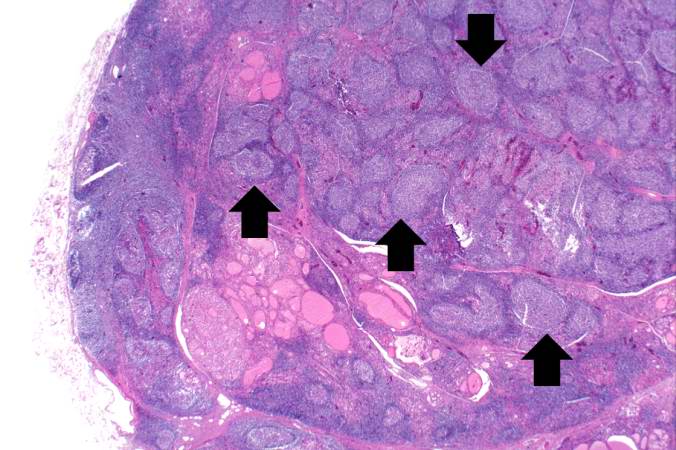
Revision as of 17:43, 20 August 2013 by Peter Anderson (talk | contribs) (This is another view of thyroid gland filled with inflammatory cells forming germinal centers (arrows).)
No higher resolution available.
IPLab6Hashimoto4.jpg (676 × 450 pixels, file size: 58 KB, MIME type: image/jpeg)
This is another view of thyroid gland filled with inflammatory cells forming germinal centers (arrows).
File history
Click on a date/time to view the file as it appeared at that time.
| Date/Time | Thumbnail | Dimensions | User | Comment | |
|---|---|---|---|---|---|
| current | 17:43, 20 August 2013 | | 676 × 450 (58 KB) | Peter Anderson (talk | contribs) | This is another view of thyroid gland filled with inflammatory cells forming germinal centers (arrows). |
- You cannot overwrite this file.
File usage
There are no pages that link to this file.