File:IPLab5PolycysticKidney6.jpg
Revision as of 17:52, 19 August 2013 by Peter Anderson (talk | contribs) (This is a higher-power photomicrograph of polycystic kidney showing the edge of a large cyst (1). In this section there are numerous tubules and dilated collecting ducts (2) that are filled with the same red proteinaceous material as the larger cysts.)
No higher resolution available.
IPLab5PolycysticKidney6.jpg (685 × 450 pixels, file size: 66 KB, MIME type: image/jpeg)
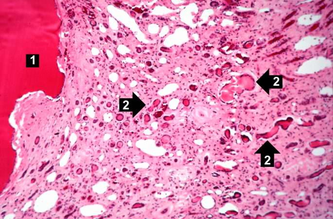
This is a higher-power photomicrograph of polycystic kidney showing the edge of a large cyst (1). In this section there are numerous tubules and dilated collecting ducts (2) that are filled with the same red proteinaceous material as the larger cysts.
File history
Click on a date/time to view the file as it appeared at that time.
| Date/Time | Thumbnail | Dimensions | User | Comment | |
|---|---|---|---|---|---|
| current | 17:52, 19 August 2013 | | 685 × 450 (66 KB) | Peter Anderson (talk | contribs) | This is a higher-power photomicrograph of polycystic kidney showing the edge of a large cyst (1). In this section there are numerous tubules and dilated collecting ducts (2) that are filled with the same red proteinaceous material as the larger cysts. |
- You cannot overwrite this file.
File usage
The following page links to this file: