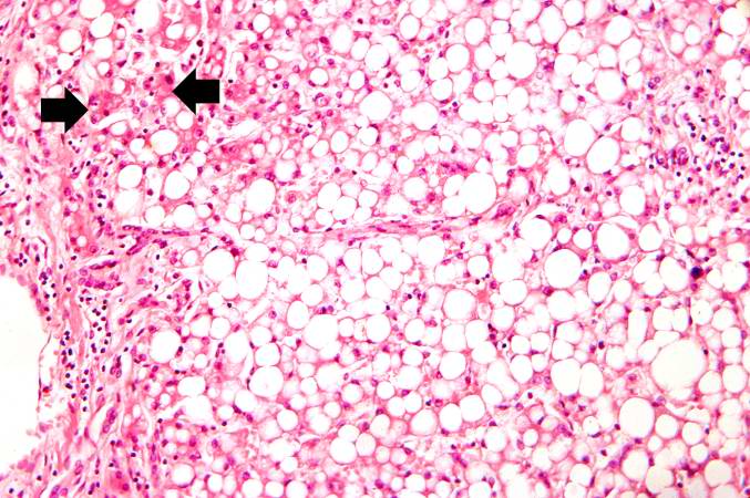
File:IPLab2FattyChange5.jpg
Revision as of 16:56, 19 August 2013 by Peter Anderson (talk | contribs) (This higher-power photomicrograph of the centrilobular area gives the appearance of fatty tissue, as indicated by many empty spaces. Very few normal liver cells can be seen in this slide. A few more normal-appearing hepatocytes are present at the left ...)
No higher resolution available.
IPLab2FattyChange5.jpg (677 × 450 pixels, file size: 75 KB, MIME type: image/jpeg)
This higher-power photomicrograph of the centrilobular area gives the appearance of fatty tissue, as indicated by many empty spaces. Very few normal liver cells can be seen in this slide. A few more normal-appearing hepatocytes are present at the left portion of the slide (arrows).
File history
Click on a date/time to view the file as it appeared at that time.
| Date/Time | Thumbnail | Dimensions | User | Comment | |
|---|---|---|---|---|---|
| current | 16:56, 19 August 2013 | | 677 × 450 (75 KB) | Peter Anderson (talk | contribs) | This higher-power photomicrograph of the centrilobular area gives the appearance of fatty tissue, as indicated by many empty spaces. Very few normal liver cells can be seen in this slide. A few more normal-appearing hepatocytes are present at the left ... |
- You cannot overwrite this file.
File usage
There are no pages that link to this file.