File:IPLab2FattyChange3.jpg
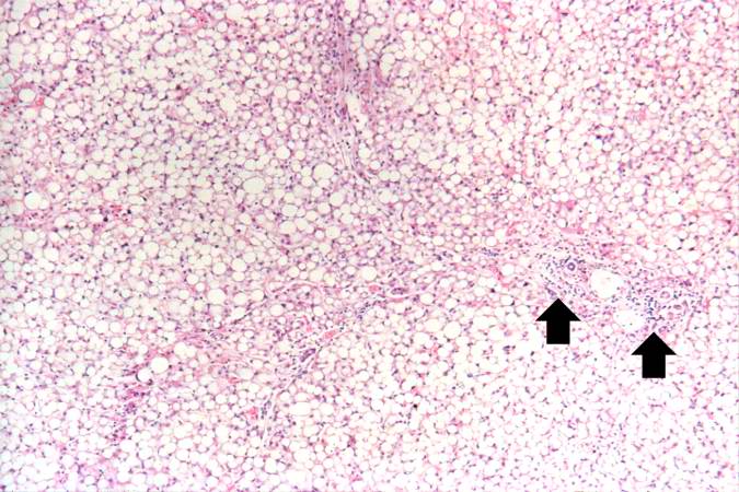
Revision as of 16:55, 19 August 2013 by Peter Anderson (talk | contribs) (Another low-power photomicrograph illustrates again the pale, washed-out appearance of this tissue. Notice the numerous holes throughout the tissue. There are accumulations of inflammatory cells (arrows) around portal tracts.)
No higher resolution available.
IPLab2FattyChange3.jpg (675 × 450 pixels, file size: 73 KB, MIME type: image/jpeg)
Another low-power photomicrograph illustrates again the pale, washed-out appearance of this tissue. Notice the numerous holes throughout the tissue. There are accumulations of inflammatory cells (arrows) around portal tracts.
File history
Click on a date/time to view the file as it appeared at that time.
| Date/Time | Thumbnail | Dimensions | User | Comment | |
|---|---|---|---|---|---|
| current | 16:55, 19 August 2013 | | 675 × 450 (73 KB) | Peter Anderson (talk | contribs) | Another low-power photomicrograph illustrates again the pale, washed-out appearance of this tissue. Notice the numerous holes throughout the tissue. There are accumulations of inflammatory cells (arrows) around portal tracts. |
- You cannot overwrite this file.
File usage
There are no pages that link to this file.