File:IPLab4Thrombosis8.jpg
Revision as of 16:40, 19 August 2013 by Seung Park (talk | contribs) (This is a higher-power photomicrograph of the vessel wall. The adventitia (1) and the media (2) contain inflammatory cells. The recanalized portion of the vessel is composed of fibrous connective tissue and contains numerous small blood vessels. There ...)
No higher resolution available.
IPLab4Thrombosis8.jpg (681 × 450 pixels, file size: 65 KB, MIME type: image/jpeg)
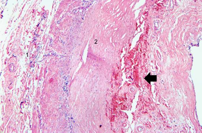
This is a higher-power photomicrograph of the vessel wall. The adventitia (1) and the media (2) contain inflammatory cells. The recanalized portion of the vessel is composed of fibrous connective tissue and contains numerous small blood vessels. There is a small area of hemorrhage (arrow) in the central portion of this image.
Recanalization is the process of the forming of channels through an organized thrombus so that blood flow is restored.
File history
Click on a date/time to view the file as it appeared at that time.
| Date/Time | Thumbnail | Dimensions | User | Comment | |
|---|---|---|---|---|---|
| current | 16:40, 19 August 2013 | | 681 × 450 (65 KB) | Seung Park (talk | contribs) | This is a higher-power photomicrograph of the vessel wall. The adventitia (1) and the media (2) contain inflammatory cells. The recanalized portion of the vessel is composed of fibrous connective tissue and contains numerous small blood vessels. There ... |
- You cannot overwrite this file.
File usage
The following page links to this file: