File:IPLab1KidneyInfarction1.jpg
Revision as of 16:17, 15 August 2013 by Seung Park (talk | contribs)
No higher resolution available.
IPLab1KidneyInfarction1.jpg (679 × 450 pixels, file size: 70 KB, MIME type: image/jpeg)
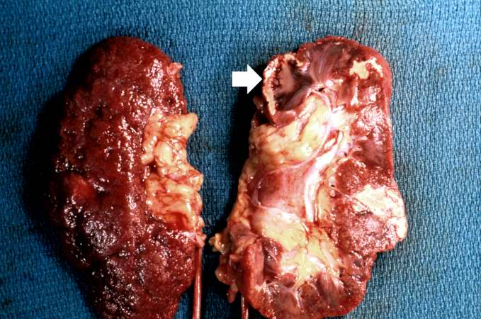
This gross photograph shows a kidney that has been transected longitudinally at autopsy. The cut surface (right) shows several areas of infarction. The most recent infarct is seen at the top left (arrow). The surface of the kidney (left) shows a marked nodularity and roughening from scarring due to chronic hypertension.
File history
Click on a date/time to view the file as it appeared at that time.
| Date/Time | Thumbnail | Dimensions | User | Comment | |
|---|---|---|---|---|---|
| current | 15:12, 15 August 2013 | | 679 × 450 (70 KB) | Seung Park (talk | contribs) |
- You cannot overwrite this file.
File usage
There are no pages that link to this file.