File:IPLab9Clostridium5.jpg
Revision as of 03:56, 21 August 2013 by Seung Park (talk | contribs) (This high-power photomicrograph shows the gas accumulation present in the tissue, a necrotic muscle cell (1), and a mild inflammatory response (2). There is also a thrombosed blood vessel (3). The blue-staining rods (bacterial organisms) can barely be ...)
No higher resolution available.
IPLab9Clostridium5.jpg (684 × 450 pixels, file size: 68 KB, MIME type: image/jpeg)
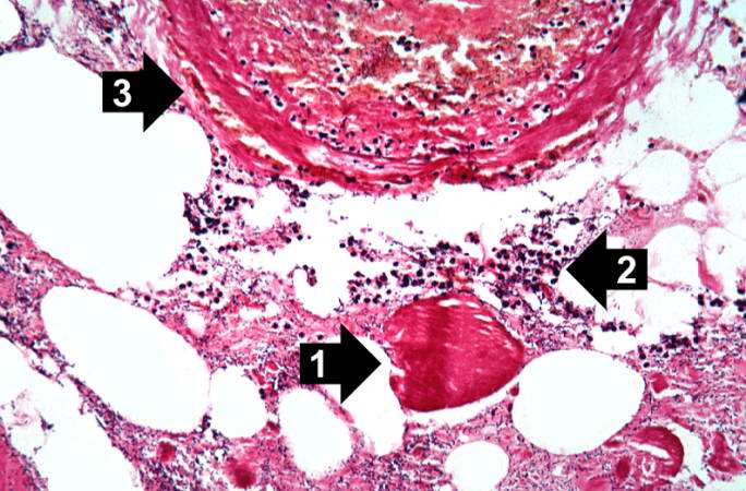
This high-power photomicrograph shows the gas accumulation present in the tissue, a necrotic muscle cell (1), and a mild inflammatory response (2). There is also a thrombosed blood vessel (3). The blue-staining rods (bacterial organisms) can barely be appreciated at this magnification.
File history
Click on a date/time to view the file as it appeared at that time.
| Date/Time | Thumbnail | Dimensions | User | Comment | |
|---|---|---|---|---|---|
| current | 03:56, 21 August 2013 | | 684 × 450 (68 KB) | Seung Park (talk | contribs) | This high-power photomicrograph shows the gas accumulation present in the tissue, a necrotic muscle cell (1), and a mild inflammatory response (2). There is also a thrombosed blood vessel (3). The blue-staining rods (bacterial organisms) can barely be ... |
- You cannot overwrite this file.
File usage
The following page links to this file: