File:IPLab9Clostridium4.jpg
Revision as of 03:56, 21 August 2013 by Seung Park (talk | contribs) (This is a high-power photomicrograph of skeletal muscle. The muscle cells are hypereosinophilic and most do not contain nuclei, indicating that these cells are dead or dying. The round clear spaces (1) in this tissue correspond to gas accumulations pri...)
No higher resolution available.
IPLab9Clostridium4.jpg (681 × 450 pixels, file size: 83 KB, MIME type: image/jpeg)
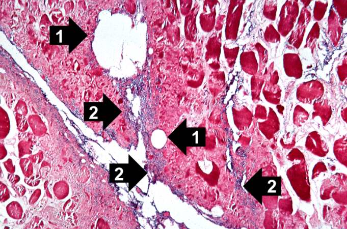
This is a high-power photomicrograph of skeletal muscle. The muscle cells are hypereosinophilic and most do not contain nuclei, indicating that these cells are dead or dying. The round clear spaces (1) in this tissue correspond to gas accumulations prior to death. In between the bundles of muscle cells, accumulations of small dark blue-staining bacterial organisms can be seen (2). Also note that there is no inflammatory response in this tissue.
File history
Click on a date/time to view the file as it appeared at that time.
| Date/Time | Thumbnail | Dimensions | User | Comment | |
|---|---|---|---|---|---|
| current | 03:56, 21 August 2013 | | 681 × 450 (83 KB) | Seung Park (talk | contribs) | This is a high-power photomicrograph of skeletal muscle. The muscle cells are hypereosinophilic and most do not contain nuclei, indicating that these cells are dead or dying. The round clear spaces (1) in this tissue correspond to gas accumulations pri... |
- You cannot overwrite this file.
File usage
The following page links to this file: