File:IPLab9BacterialMeningitis5.jpg
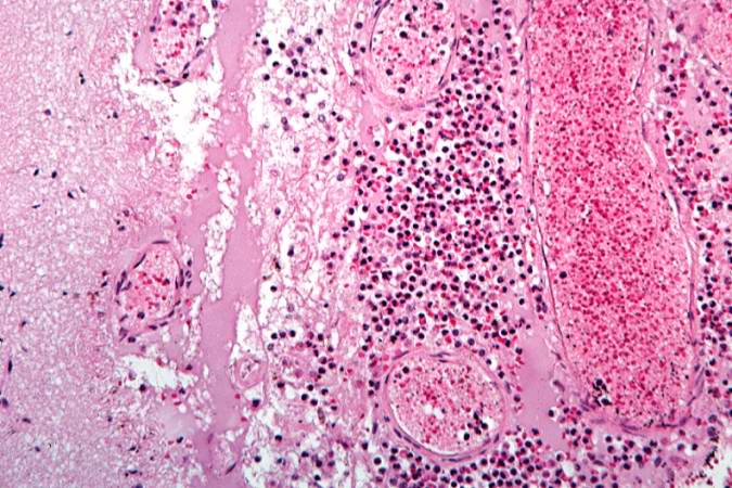
Revision as of 03:47, 21 August 2013 by Seung Park (talk | contribs) (This is a higher-power photomicrograph of inflammatory exudate in a sulcus. The majority of cells in this exudate are neutrophils. There is also abundant fibrin (arrows) and red blood cells are present in the congested vessels.)
No higher resolution available.
IPLab9BacterialMeningitis5.jpg (675 × 450 pixels, file size: 78 KB, MIME type: image/jpeg)
This is a higher-power photomicrograph of inflammatory exudate in a sulcus. The majority of cells in this exudate are neutrophils. There is also abundant fibrin (arrows) and red blood cells are present in the congested vessels.
File history
Click on a date/time to view the file as it appeared at that time.
| Date/Time | Thumbnail | Dimensions | User | Comment | |
|---|---|---|---|---|---|
| current | 03:47, 21 August 2013 | | 675 × 450 (78 KB) | Seung Park (talk | contribs) | This is a higher-power photomicrograph of inflammatory exudate in a sulcus. The majority of cells in this exudate are neutrophils. There is also abundant fibrin (arrows) and red blood cells are present in the congested vessels. |
- You cannot overwrite this file.
File usage
The following page links to this file: