File:IPLab8HSVGlossitis3.jpg
Revision as of 02:29, 21 August 2013 by Seung Park (talk | contribs) (This is a medium-power photomicrograph of epithelium at the edge of the ulcer. Even at this power, amphophilic (dark, blue-purple-staining) intranuclear inclusion bodies can be seen in the epithelial cells. Note the inflammatory infiltrate in the subep...)
No higher resolution available.
IPLab8HSVGlossitis3.jpg (673 × 450 pixels, file size: 75 KB, MIME type: image/jpeg)
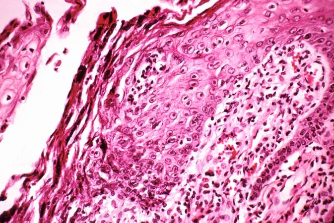
This is a medium-power photomicrograph of epithelium at the edge of the ulcer. Even at this power, amphophilic (dark, blue-purple-staining) intranuclear inclusion bodies can be seen in the epithelial cells. Note the inflammatory infiltrate in the subepithelium.
An infiltrate is an accumulation of cells in the lung parenchyma--this is a sign of pneumonia.
File history
Click on a date/time to view the file as it appeared at that time.
| Date/Time | Thumbnail | Dimensions | User | Comment | |
|---|---|---|---|---|---|
| current | 02:29, 21 August 2013 | | 673 × 450 (75 KB) | Seung Park (talk | contribs) | This is a medium-power photomicrograph of epithelium at the edge of the ulcer. Even at this power, amphophilic (dark, blue-purple-staining) intranuclear inclusion bodies can be seen in the epithelial cells. Note the inflammatory infiltrate in the subep... |
- You cannot overwrite this file.
File usage
The following page links to this file: