File:IPLab8HSVGlossitis1.jpg
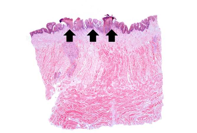
Revision as of 02:28, 21 August 2013 by Seung Park (talk | contribs) (This is a low-power photomicrograph showing a cross section of the tongue. There is an area along the surface of the tongue where the normal epithelium has been lost and there are areas of ulceration (arrows).)
No higher resolution available.
IPLab8HSVGlossitis1.jpg (668 × 450 pixels, file size: 38 KB, MIME type: image/jpeg)
This is a low-power photomicrograph showing a cross section of the tongue. There is an area along the surface of the tongue where the normal epithelium has been lost and there are areas of ulceration (arrows).
File history
Click on a date/time to view the file as it appeared at that time.
| Date/Time | Thumbnail | Dimensions | User | Comment | |
|---|---|---|---|---|---|
| current | 02:28, 21 August 2013 | | 668 × 450 (38 KB) | Seung Park (talk | contribs) | This is a low-power photomicrograph showing a cross section of the tongue. There is an area along the surface of the tongue where the normal epithelium has been lost and there are areas of ulceration (arrows). |
- You cannot overwrite this file.
File usage
The following page links to this file: