File:IPLab7Carcinoid5.jpg
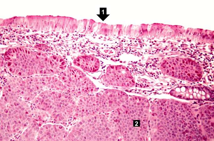
Revision as of 02:07, 21 August 2013 by Seung Park (talk | contribs) (This is a low-power photomicrograph of one of the subcutaneous masses in the cecum. Note that the mucosa (1) is virtually normal and the tumor cells are in the submucosa (2).)
No higher resolution available.
IPLab7Carcinoid5.jpg (683 × 450 pixels, file size: 65 KB, MIME type: image/jpeg)
This is a low-power photomicrograph of one of the subcutaneous masses in the cecum. Note that the mucosa (1) is virtually normal and the tumor cells are in the submucosa (2).
File history
Click on a date/time to view the file as it appeared at that time.
| Date/Time | Thumbnail | Dimensions | User | Comment | |
|---|---|---|---|---|---|
| current | 02:07, 21 August 2013 | | 683 × 450 (65 KB) | Seung Park (talk | contribs) | This is a low-power photomicrograph of one of the subcutaneous masses in the cecum. Note that the mucosa (1) is virtually normal and the tumor cells are in the submucosa (2). |
- You cannot overwrite this file.
File usage
There are no pages that link to this file.