File:IPLab7Melanoma5.jpg
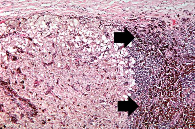
Revision as of 01:56, 21 August 2013 by Seung Park (talk | contribs) (This is a higher-magnification showing the abundant extracellular melanin (arrows) surrounding the tumor cells. This section of neoplasm shows the numerous cells with abundant cytoplasm and brown pigment within the cytoplasm of some of these cells.)
No higher resolution available.
IPLab7Melanoma5.jpg (679 × 450 pixels, file size: 93 KB, MIME type: image/jpeg)
This is a higher-magnification showing the abundant extracellular melanin (arrows) surrounding the tumor cells. This section of neoplasm shows the numerous cells with abundant cytoplasm and brown pigment within the cytoplasm of some of these cells.
File history
Click on a date/time to view the file as it appeared at that time.
| Date/Time | Thumbnail | Dimensions | User | Comment | |
|---|---|---|---|---|---|
| current | 01:56, 21 August 2013 | | 679 × 450 (93 KB) | Seung Park (talk | contribs) | This is a higher-magnification showing the abundant extracellular melanin (arrows) surrounding the tumor cells. This section of neoplasm shows the numerous cells with abundant cytoplasm and brown pigment within the cytoplasm of some of these cells. |
- You cannot overwrite this file.
File usage
There are no pages that link to this file.