File:IPLab7Melanoma4.jpg
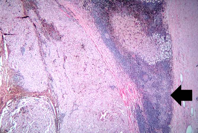
Revision as of 01:56, 21 August 2013 by Seung Park (talk | contribs) (This higher-power photomicrograph shows the remaining portion of lymph node (arrow). The rest of the lymph node is invaded by a neoplasm composed of cells with lighter eosinophilic cytoplasm and pigment.)
No higher resolution available.
IPLab7Melanoma4.jpg (669 × 450 pixels, file size: 74 KB, MIME type: image/jpeg)
This higher-power photomicrograph shows the remaining portion of lymph node (arrow). The rest of the lymph node is invaded by a neoplasm composed of cells with lighter eosinophilic cytoplasm and pigment.
File history
Click on a date/time to view the file as it appeared at that time.
| Date/Time | Thumbnail | Dimensions | User | Comment | |
|---|---|---|---|---|---|
| current | 01:56, 21 August 2013 | | 669 × 450 (74 KB) | Seung Park (talk | contribs) | This higher-power photomicrograph shows the remaining portion of lymph node (arrow). The rest of the lymph node is invaded by a neoplasm composed of cells with lighter eosinophilic cytoplasm and pigment. |
- You cannot overwrite this file.
File usage
There are no pages that link to this file.