File:IPLab7IDC1.jpg
Revision as of 01:50, 21 August 2013 by Seung Park (talk | contribs) (This is a gross photograph of the surgical specimen of breast with infiltrating duct carcinoma. Note the tumor tissue under the area of the nipple. The tumor infiltrates in an irregular fashion into the breast parenchyma. Note the nipple retraction cau...)
No higher resolution available.
IPLab7IDC1.jpg (722 × 450 pixels, file size: 56 KB, MIME type: image/jpeg)
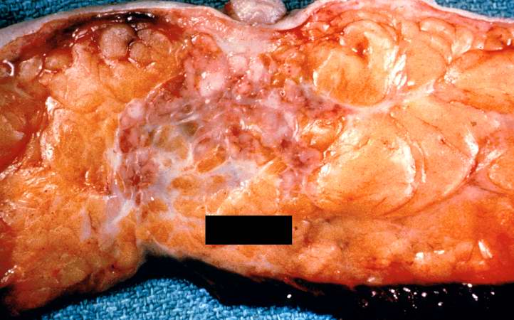
This is a gross photograph of the surgical specimen of breast with infiltrating duct carcinoma. Note the tumor tissue under the area of the nipple. The tumor infiltrates in an irregular fashion into the breast parenchyma. Note the nipple retraction caused by this neoplasm.
An infiltrate is an accumulation of cells in the lung parenchyma--this is a sign of pneumonia.
File history
Click on a date/time to view the file as it appeared at that time.
| Date/Time | Thumbnail | Dimensions | User | Comment | |
|---|---|---|---|---|---|
| current | 01:50, 21 August 2013 | | 722 × 450 (56 KB) | Seung Park (talk | contribs) | This is a gross photograph of the surgical specimen of breast with infiltrating duct carcinoma. Note the tumor tissue under the area of the nipple. The tumor infiltrates in an irregular fashion into the breast parenchyma. Note the nipple retraction cau... |
- You cannot overwrite this file.
File usage
There are no pages that link to this file.