File:IPLab7Fibroadenoma4.jpg
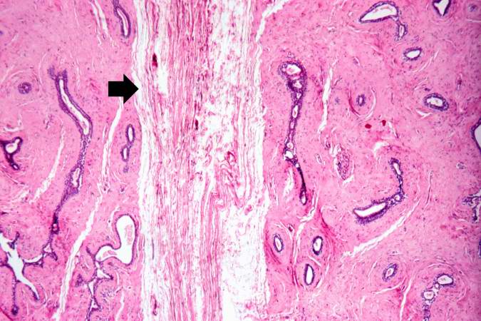
Revision as of 01:27, 21 August 2013 by Seung Park (talk | contribs) (This photomicrograph shows the compressed connective tissue (arrow) between two nodules of dense fibrous tissue and ducts.)
No higher resolution available.
IPLab7Fibroadenoma4.jpg (675 × 450 pixels, file size: 67 KB, MIME type: image/jpeg)
This photomicrograph shows the compressed connective tissue (arrow) between two nodules of dense fibrous tissue and ducts.
File history
Click on a date/time to view the file as it appeared at that time.
| Date/Time | Thumbnail | Dimensions | User | Comment | |
|---|---|---|---|---|---|
| current | 01:27, 21 August 2013 | | 675 × 450 (67 KB) | Seung Park (talk | contribs) | This photomicrograph shows the compressed connective tissue (arrow) between two nodules of dense fibrous tissue and ducts. |
- You cannot overwrite this file.
File usage
There are no pages that link to this file.