File:IPLab6ChronicRejection10.jpg
Revision as of 21:50, 20 August 2013 by Seung Park (talk | contribs) (This is a high-power photomicrograph of a kidney from another case of chronic transplant rejection. In this case there is extensive damage to the kidney due to the chronic rejection (loss of tubules and glomerular lesions). In addition, this kidney was...)
No higher resolution available.
IPLab6ChronicRejection10.jpg (684 × 450 pixels, file size: 86 KB, MIME type: image/jpeg)
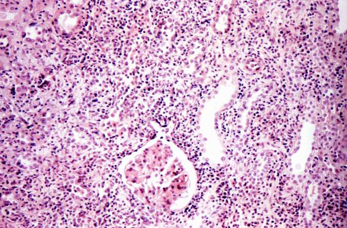
This is a high-power photomicrograph of a kidney from another case of chronic transplant rejection. In this case there is extensive damage to the kidney due to the chronic rejection (loss of tubules and glomerular lesions). In addition, this kidney was removed during an episode of acute rejection. The marked cellular infiltrate indicates acute rejection in a case of chronic transplant rejection.
An infiltrate is an accumulation of cells in the lung parenchyma--this is a sign of pneumonia.
File history
Click on a date/time to view the file as it appeared at that time.
| Date/Time | Thumbnail | Dimensions | User | Comment | |
|---|---|---|---|---|---|
| current | 21:50, 20 August 2013 | | 684 × 450 (86 KB) | Seung Park (talk | contribs) | This is a high-power photomicrograph of a kidney from another case of chronic transplant rejection. In this case there is extensive damage to the kidney due to the chronic rejection (loss of tubules and glomerular lesions). In addition, this kidney was... |
- You cannot overwrite this file.
File usage
The following page links to this file: