File:IPLab6ChronicRejection7.jpg
Revision as of 21:49, 20 August 2013 by Seung Park (talk | contribs) (This is a photomicrograph of a glomerulus with a mild cellular infiltrate (left) and a small damaged glomerulus (right). There is extensive interstitial fibrosis (1), loss of renal tubules, and the remaining tubules contain protein (2) indicating sever...)
No higher resolution available.
IPLab6ChronicRejection7.jpg (676 × 450 pixels, file size: 70 KB, MIME type: image/jpeg)
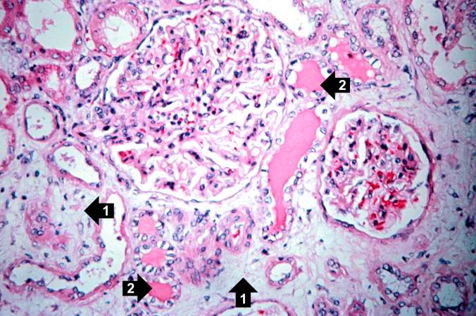
This is a photomicrograph of a glomerulus with a mild cellular infiltrate (left) and a small damaged glomerulus (right). There is extensive interstitial fibrosis (1), loss of renal tubules, and the remaining tubules contain protein (2) indicating severe damage.
An infiltrate is an accumulation of cells in the lung parenchyma--this is a sign of pneumonia.
File history
Click on a date/time to view the file as it appeared at that time.
| Date/Time | Thumbnail | Dimensions | User | Comment | |
|---|---|---|---|---|---|
| current | 21:49, 20 August 2013 | | 676 × 450 (70 KB) | Seung Park (talk | contribs) | This is a photomicrograph of a glomerulus with a mild cellular infiltrate (left) and a small damaged glomerulus (right). There is extensive interstitial fibrosis (1), loss of renal tubules, and the remaining tubules contain protein (2) indicating sever... |
- You cannot overwrite this file.
File usage
The following page links to this file: