File:IPLab6Amyloid6.jpg
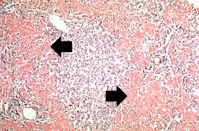
Revision as of 21:35, 20 August 2013 by Seung Park (talk | contribs) (This is a low-power photomicrograph of liver tissue stained with Congo red (orange color in slide). Congo red reacts with amyloid and gives it an orange color (arrows).)
No higher resolution available.
IPLab6Amyloid6.jpg (685 × 450 pixels, file size: 92 KB, MIME type: image/jpeg)
This is a low-power photomicrograph of liver tissue stained with Congo red (orange color in slide). Congo red reacts with amyloid and gives it an orange color (arrows).
File history
Click on a date/time to view the file as it appeared at that time.
| Date/Time | Thumbnail | Dimensions | User | Comment | |
|---|---|---|---|---|---|
| current | 21:35, 20 August 2013 | | 685 × 450 (92 KB) | Seung Park (talk | contribs) | This is a low-power photomicrograph of liver tissue stained with Congo red (orange color in slide). Congo red reacts with amyloid and gives it an orange color (arrows). |
- You cannot overwrite this file.
File usage
The following page links to this file: