File:IPLab5Gout3.jpg
Revision as of 15:18, 20 August 2013 by Peter Anderson (talk | contribs) (This is a low-power photomicrograph of the tophus removed from the elbow of this patient. Note the fibrous connective tissue (1) and the large foci containing the urate crystals (2) surrounded by the intense chronic inflammatory reaction.)
No higher resolution available.
IPLab5Gout3.jpg (634 × 450 pixels, file size: 21 KB, MIME type: image/jpeg)
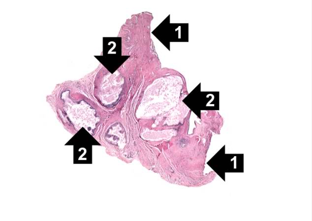
This is a low-power photomicrograph of the tophus removed from the elbow of this patient. Note the fibrous connective tissue (1) and the large foci containing the urate crystals (2) surrounded by the intense chronic inflammatory reaction.
A tophus is a chalky accumulation of urate crystals found in the tissue surrounding a joint.
File history
Click on a date/time to view the file as it appeared at that time.
| Date/Time | Thumbnail | Dimensions | User | Comment | |
|---|---|---|---|---|---|
| current | 15:18, 20 August 2013 | | 634 × 450 (21 KB) | Peter Anderson (talk | contribs) | This is a low-power photomicrograph of the tophus removed from the elbow of this patient. Note the fibrous connective tissue (1) and the large foci containing the urate crystals (2) surrounded by the intense chronic inflammatory reaction. |
- You cannot overwrite this file.
File usage
The following page links to this file: