File:IPLab6GravesDisease2.jpg
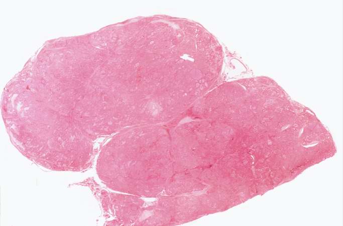
Revision as of 18:22, 19 August 2013 by Seung Park (talk | contribs) (This is a low-power photomicrograph of thyroid tissue from this case. The tissue is very cellular with very little colloid.)
No higher resolution available.
IPLab6GravesDisease2.jpg (683 × 450 pixels, file size: 29 KB, MIME type: image/jpeg)
This is a low-power photomicrograph of thyroid tissue from this case. The tissue is very cellular with very little colloid.
File history
Click on a date/time to view the file as it appeared at that time.
| Date/Time | Thumbnail | Dimensions | User | Comment | |
|---|---|---|---|---|---|
| current | 18:22, 19 August 2013 | | 683 × 450 (29 KB) | Seung Park (talk | contribs) | This is a low-power photomicrograph of thyroid tissue from this case. The tissue is very cellular with very little colloid. |
- You cannot overwrite this file.
File usage
The following page links to this file: